Introduction to Bena Village
Bena lies at the eastern foot of the Inerie volcano, which is 2,245 metres high, in the Ngada region of the Indonesian Island of Flores, between ten and twenty kilometers south of the town of Bajawa. The full name of the village is Kampung Adat Bena, which means „Bena traditional village” in Indonesian. Sources also use the name Kampung Tradisional Bena. In the administrative system, Bena belongs to the village of Tiworiwu in the Jerebu’u district, in the Ngada regency, in the province of East Nusa Tenggara (Nusa Tenggara Timur).
Bena is the oldest village of the Ngada people. According to the village elders, the village has never moved from its original site. The age of the village is estimated at about 1,200 years. Bena stretches from north to south along the crest of a hill. Nine clans live here in 45 houses, which stand on terraces that step down the slope. The traditional house of the Ngada people is called sa’o: a steep thatched roof (a roof covered with dry grass) on a wooden structure.
The community is matrilineal: family descent is counted through the mother’s line. The Bena clan, regarded as the founders of the village, occupies the middle terrace.
From the ground, Bena is simply a village. Two rows of houses, an open yard in the middle, steep roofs, ancestral stones. A visitor enters at the lower end, walks up along one row of houses and walks back down along the other row.
A view from the air shows something different.
A closed pouch. One layer of houses around an empty central space. A dead end on one side. On the other side, a narrowing, and after the narrowing, a single exit that turns into a road.
My description of the village is based on written sources and on drone and satellite photographs. I have not visited Bena. Everything I read in the following sections is visible from above.
To see what Bena shows, we first need to know what we are looking for.
The Place We Are Looking For: Bena and the Pancreas
Illustration of pancreas and pancreatic islet cells. BlueRingMedia – Shutterstock.
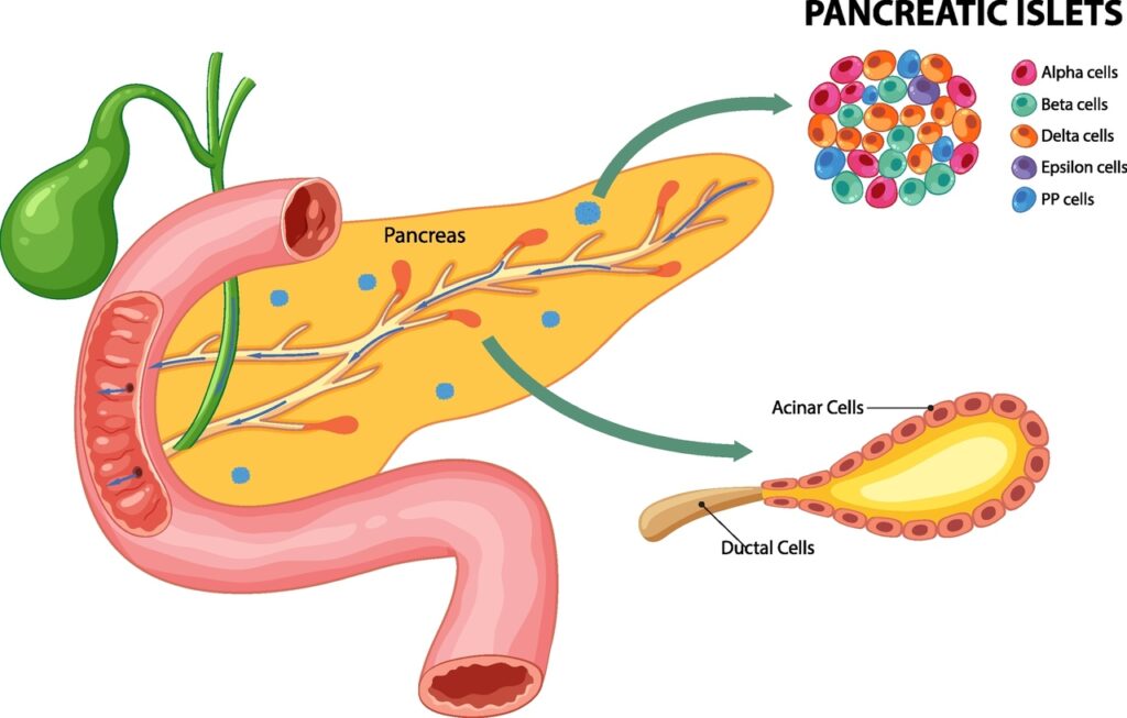
The pancreas lies in the abdomen, behind the stomach. The head of the pancreas (its widest part) rests in the curve of the duodenum, the first section of the small intestine. The pancreas is two glands in one organ.
The first gland consists of the pancreatic islets (the islets of Langerhans). In the illustration, the islets are the small blue clusters scattered across the organ. The islets have no ducts. The islets release hormones directly into the blood. This part of the organ is the endocrine pancreas (the hormone-producing pancreas). The human pancreas contains between one and two million islets, and together the islets make up only 1–2 percent of the mass of the organ.
The illustration enlarges one blue dot. A pancreatic islet is not uniform. A pancreatic islet is a cluster of five types of cells, mixed together. Each type of islet cell produces a different hormone:
Beta cells – the most numerous islet cells; in humans, beta cells make up more than half of the cells in an islet. Beta cells produce insulin, the hormone that lowers the level of sugar in the blood and allows the body’s cells to take in glucose. Beta cells are the only cells in the body that produce insulin. When the immune system destroys the beta cells, type 1 diabetes develops.
(PDB 1MSO)

Alpha cells – the second most numerous group. Alpha cells produce glucagon, a hormone with the opposite action to insulin. When the blood has too little sugar, glucagon signals the liver to release its stored sugar.
(PDB 1GCN)

Delta cells – a few percent of the cells in an islet. Delta cells produce somatostatin, an inhibiting hormone (a hormone that slows other cells down). Somatostatin reduces the release of insulin and glucagon by the neighbouring cells. Somatostatin acts as a local brake.
(PDB 7T10)

PP cells – few in number, and most common in the head of the pancreas. PP cells produce pancreatic polypeptide. Pancreatic polypeptide reduces the release of digestive fluid by the acini and influences appetite. Pancreatic polypeptide is one of the links between the islets and the acini.
(PDB 7X9C)

Epsilon cells – the rarest islet cells of all. Epsilon cells produce ghrelin, the hunger hormone.
(PDB 7F9Y)

Two hormones lead the main process: insulin lowers blood sugar, and glucagon raises blood sugar. Somatostatin makes sure that neither insulin nor glucagon goes too far. Pancreatic polypeptide and ghrelin connect this process to digestion and to hunger.
Five types of cells. Five hormones. One cluster the size of a grain of sand that keeps the blood sugar of the whole human body in balance.
The second gland is all the rest of the organ: the exocrine pancreas (the part of the pancreas that releases its product into ducts). The exocrine pancreas produces pancreatic juice: enzymes that digest proteins, fats and carbohydrates, and a fluid rich in bicarbonate, which neutralises the acid that arrives from the stomach. Every day the pancreas produces litres of this juice.
Pancreatic juice is not made in the ducts. Pancreatic juice is made at the ends of the ducts.
In the illustration, the ducts branch like a tree. The smallest branches end in red drops. Each red drop is a secretory acinus, from the Latin word acinus. The Latin word means a berry, a grape. The acini hang from the ducts like grapes hanging from the stems of a grape cluster.
The ducts collect pancreatic juice from thousands of acini. Small branches join into larger branches, and larger branches join into the main pancreatic duct. Just before the intestine, the green common bile duct joins the main pancreatic duct. The common bile duct carries bile from the liver and the gallbladder. Both ducts open together into the duodenum. Only in the duodenum do the enzymes begin to work.
The enlargement on the right of the illustration shows one acinus.
An acinus is a pouch with walls made of a single layer of cells. The walls are formed by acinar cells (the secretory cells of the acinus), placed side by side in a closed ring. In the middle of the ring there is an empty space: the lumen of the acinus (the central space inside the acinus). The acinar cells release their product into the lumen.
Each acinar cell has two sides. The wide base of the cell (the basal side) faces outward, towards the blood vessels. The nucleus and the protein factory of the cell are located on the basal side. The narrow tip of the cell (the apical side) faces the lumen. Finished secretory granules (small storage packages) filled with enzymes wait on the apical side. The acinar cell works in one direction: from the base to the tip, from the outside to the inside.
The enzymes leave the acinar cell in an inactive form. If the enzymes became active inside the acinus, the pancreas would digest itself. The enzymes become active only in the duodenum.
The acinus has one closed end. The other end narrows into a neck (a narrow passage). After the neck, ductal cells take over the walls; ductal cells are lower and flatter than acinar cells. This is where the intercalated duct begins: the smallest branch of the tree of ducts.
One point requires honesty. In the illustration, the lumen of the acinus is wide. In a real pancreas, the lumen is a narrow gap. Diagrams make the lumen larger to show where the secretion goes. Bena matches the diagram. This is the silhouette level of reading (the outline of the shape, seen from a distance).
A closed pouch. One layer of cells around a central lumen. A neck. A duct.
This is what we are looking for.
Let us begin to read the village from its shape.
The Mapping – Bena Village and the Pancreatic Acinus
image description: Bird’s-eye view of Bena Traditional houses in the traditional village of Bena on the island of Flores in Indonesia. Lee Risar – Shutterstock.
Vector description: Detailed view of acinar and ductal cells. BlueRingMedia – Shutterstock.
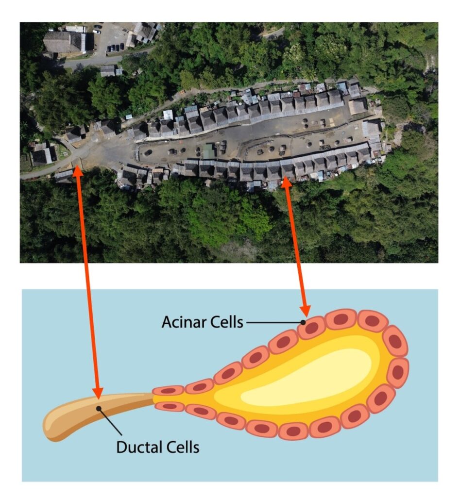
The photograph and the diagram have the same orientation. The exit is on the left. The closed end is on the right. Nothing needs to be rotated.
The Acinar Cells of the Pancreas = The Houses in Two Rows
The right-hand red arrow connects a house in the lower row with a cell in the wall of the acinus.
The houses stand side by side. Roof next to roof, with no gaps. The houses form two rows, an upper row and a lower row, which run along the whole length of the village. Each row is a single layer. There is no second line of houses behind the first line.
In the diagram, the acinar cells form a single layer in the wall of the acinus. The acinar cells also stand side by side. There is also no second line of cells.
Each house has a steep roof with four sloping sides. From above, each roof looks like a pyramid. Histology describes acinar cells as pyramidal cells (cells shaped like a pyramid).
The Lumen of the Acinus = The Central Village Yard
Between the two rows of houses lies a long open yard. The front of every house faces the yard, on both sides.
In the diagram, a yellow lumen lies between the walls of the acinus. The tip of every acinar cell faces the lumen.
The village yard is not empty. Round and rectangular objects stand along the whole length of the yard. In the diagram, the lumen is not empty either: secretion fills the lumen. The meaning of these objects belongs to the next level of reading. The drone photograph shows only that the objects are there – in the middle, between the two rows of houses.
The Closed End of the Acinus = The Right-Hand End of the Village
On the right-hand side, the rows of houses do not end with an opening. Buildings placed across the end of the village close the rows. The yard has no exit at this end.
In the diagram, the right-hand end of the acinus is rounded and closed. The wall of cells continues from the upper side to the lower side without a gap.
A dead end. A pouch.
The Ductal Cells = The Village Exit and the Road
The left-hand red arrow connects the road leaving the village with the duct in the diagram.
On the left-hand side, the rows of houses end. The space of the yard opens into open ground, and the open ground turns into a road. The road continues to the left and leaves the frame of the photograph.
In the diagram, the acinus narrows into a neck, and the neck turns into a duct. The duct continues to the left, away from the acinus.
One entrance. One exit. The same place.
Near the left-hand end, especially in the lower row, the houses are smaller and stand less regularly than in the middle of the village. In the diagram, the acinar cells near the neck are also smaller and lower than the acinar cells at the closed end. After these smaller cells, the flat ductal cells begin.
The Basal Side of the Acinar Cell = The Back of the Houses
At the back of the houses, on the side facing the forest, there are small extensions with light metal roofs. The front of each house faces the yard. The back of each house faces outward.
An acinar cell also has two sides. The tip of the cell faces the lumen. The base of the cell faces outward, and the protein factory of the cell works at the base. In a real acinar cell, the nucleus also lies near the base. The diagram does not show this detail, because the diagram draws each nucleus in the middle of the cell.
The Surrounding Tissue = The Forest
Forest surrounds the village. The forest reaches directly to the back walls of the houses, on both sides. There is no wall and no ring road between the forest and the houses.
In the diagram, the acinus lies on a blue background. In the pancreas, this background is connective tissue with blood vessels. A pancreatic acinus has no extra layer of contractile cells (muscle-like cells that squeeze the gland) around its wall; acini in the salivary glands do have such a layer. The wall of acinar cells touches the surrounding tissue directly.
The houses of Bena touch the forest in the same way.
The shape of a pouch. One layer of cells. A lumen filled with secretion. A dead end. A neck and a duct. Two different sides of each cell. Surrounding tissue with no outer ring.
The pattern matches.
Proteins in the Living Cell = The Residents of Bena
People are not visible from the drone. Proteins are not visible in the diagram of the acinus. Yet the proteins do all the work.
The houses are the cells. The residents are the proteins: the molecular workers that perform actions inside the living cell. Proteins build, carry, cut, connect and repair. A cell without proteins is an empty house.
Nine clans live in the 45 houses of Bena. Men and women work mainly in the fields. Women weave ikat cloth (a traditional dyed and woven textile). The work takes place in and around the houses, and the products of the work travel further.
An acinar cell works in the same way. Inside the acinar cell, some proteins maintain the cell: these proteins fold new protein chains, pack the chains and keep order. Biology calls some of these proteins resident proteins (proteins that stay inside the cell). Resident proteins stay in place and work for the cell.
Other proteins are made in order to leave the cell. These proteins are the digestive enzymes. The acinar cell produces the digestive enzymes, packs the enzymes into granules at the tip of the cell and releases the enzymes into the lumen of the acinus. From the lumen, the enzymes flow through the duct to the intestine. In the intestine, the enzymes do their work, far from the cell in which the enzymes were made.
Bena has its residents. The residents are the people who stay in the village and maintain the houses, the clans and the ceremonies. Bena also has people who left. Thousands of descendants of Bena’s inhabitants live outside the village today, and these descendants remain connected to the village through tradition. The descendants left along the only road, through the neck of the acinus, through the duct. The descendants work in the wider world, far from the house in which they were born.
Resident proteins and secreted proteins. Villagers who stay and villagers who leave.
One living cell. One village. The same division of work.
Two Acini on One Branch of the Duct – Bena and the Smaller Settlement
Bena is not alone.
Image description: Aerial view of the ancient Bena Village in Bajawa, Flores, Indonesia. Beautiful landscape of the thatched roof village in a calm and contemplating morning ambience surrounded by hills and cliffs. Lee Risar – Shutterstock.
Vector description: Detailed view of acinar and ductal cells. BlueRingMedia – Shutterstock.

The upper image is the same drone photograph as at the beginning of this article, rotated. In the rotated photograph, the roads leave the villages to the left – in the same direction in which pancreatic juice flows in the illustration: to the left, towards the duodenum.
Bena lies in the middle of the frame. A long, narrow pouch. Two rows of houses, an open yard in the middle.
In the upper left corner, above the forest, lies a second settlement. The second settlement is smaller. The second settlement also has houses around a central yard, with the front of each house facing the middle. On maps, the smaller settlement has no name of its own.
Both settlements have the same layout. Houses in a single layer around a yard. The yard in the middle. The exit in one place.
The two settlements differ in size and shape. Bena is long and narrow. The smaller settlement is short and compact, almost square. The smaller settlement also has more light metal roofs.
Now the roads.
A short road leaves the smaller settlement. The road goes down towards the exit of Bena. A second road leaves Bena at its left-hand end. Both roads meet on the left-hand side of the frame. From that point, the two roads continue as one road.
Two settlements. Two short roads. One shared road.
An orange wedge-shaped pointer leads from the photograph to the illustration of the pancreas. The pointer marks one place in the tree of ducts: the point where small branches carrying acini join into a larger branch.
The ducts of the pancreas branch like a tree. An acinus hangs at the end of each smallest branch. The smallest branch is the intercalated duct, the first and shortest section of the path along which pancreatic juice flows. Several intercalated ducts join into a larger duct. In this way, the acini gather into clusters, like grapes.
The acini in one cluster are not identical. Some acini are long, other acini are round. Some acini are larger, other acini are smaller. The acini share the same structure and a shared exit.
Bena and the smaller settlement show exactly this part of the illustration. Two acini of different sizes. Each acinus has its own intercalated duct. Both intercalated ducts join into one duct.
Acinus means berry.
Two berries on one grape stem.
An Acinus with Missing Cells – Missing Houses and Their Foundations
Image desription: Aerial view of traditional house of „Ratenggaro – Sumba, East Nusa Tenggara” Indonesia. MawardiBahar – Shutterstock.
Vector description: Detailed view of acinar and ductal cells. BlueRingMedia – Shutterstock.

This photograph shows another village from the same region of Indonesia. The houses are of the same type: a steep, pyramid-shaped roof covered with thatch. The diagram below the photograph is the same as before.
The Same Layout of the Acinus, More Loosely Arranged
The houses stand on both sides of a narrow space in the middle. The houses run diagonally across the frame, from the upper left corner to the lower right corner. The front of each house faces the middle.
The right-hand red arrow connects a house in the lower part of the village with a cell in the wall of the acinus. The left-hand red arrow connects the road with the duct in the diagram; the road leaves the village in the upper left corner and continues upward in the frame.
Near the exit to the road, the houses are smaller and stand further apart. This is the same as in Bena. This is the same as the acinar cells near the neck of the acinus.
The rows are not as tightly closed here as in Bena. There are gaps between the houses.
The Roof of a House as a Cell: Light Thatch and a Dark Nucleus
In this photograph, the light falls in a way that shows each roof separately.
Each roof has the shape of a pyramid. The thatch on the slopes of the roof is light, the colour of sand. In the very centre, at the top of the roof, the colour becomes darker. A dark, pointed centre. A light area all around the centre.
Look at the diagram below the photograph. Each acinar cell is a light shape. A dark oval lies in the middle of each light shape. The dark oval is the cell nucleus, the place where the genome is stored.
Light all around. Dark in the centre. The roof is the cell. The dark top of the roof is the cell nucleus.
Nothing needs to be added. It is enough to place the photograph next to the diagram.
One reservation. The diagram draws the nucleus in the middle of the cell, and the roofs repeat this picture. In a living acinar cell, the nucleus is shifted towards the base of the cell, because the enzyme granules fill the tip of the cell. The roofs match the diagram. This is still the silhouette level of reading.
Gaps in the Wall of the Acinus
The gaps are not empty.
In several places between the houses, there are bare rectangles of earth and stone. Each rectangle has the shape and size of a house. The rectangles lie in line with the row, where the next house should stand. The house is missing. The plan of the building remains, written in stone.
In the middle part of the village, in one of these gaps, stands a light lattice frame (a grid of poles). The frame has the size of a house, but the frame has no roof and no walls. The drone photograph does not show whether the frame is the skeleton of a new house or something else.
The photograph does not explain why the houses are missing. The photograph shows only that their places remain.
The Acinar Cell That Is Missing
The acinar cells of the pancreas live for a long time, but not forever. A damaged or worn-out acinar cell goes through apoptosis, which is programmed cell death. The cell breaks down in an orderly way, and cleaning cells remove the remains. A gap remains in the wall of the acinus.
The gap does not disappear without a trace. Each acinar cell stands on a basement membrane, a thin layer of proteins that separates the lining tissue of the gland from the connective tissue. When the acinar cell dies, the basement membrane remains. The basement membrane keeps the shape and the position of the cell that is gone.
A foundation without a house.
The wall of the acinus is rebuilt on this support structure. Studies in mice have shown that new acinar cells are formed mainly by the division of surviving neighbouring acinar cells. A neighbouring cell divides, and the new cell takes the empty place. The new cell fits into the same plan that the previous cell left behind.
If the lattice frame in the middle of the village is the skeleton of a new house, the frame stands exactly where it should stand. On the old foundation. In line with the row.
Two Acini, Two States
Bena is a complete acinus. The wall is closed, house next to house.
The second village is an acinus during renewal. Some cells stand. Some cells are missing. The places of the missing cells are waiting.
Every pancreas knows both states. The wall of an acinus is not given once and for all. The wall of an acinus is maintained.
Conclusions – What the Pancreatic Acinus Teaches
The Acinar Cell That Taught Science How Cells Secrete Proteins
The exocrine pancreas is the organ with the highest level of protein production in the adult body. The acinar cells of the pancreas produce and release more protein than any other type of cell in an adult human. Most of what the acinar cells produce, the acinar cells send outside the cell.
For this reason, science first traced the secretory pathway (the route that proteins follow from production to release) in these cells. In the years 1950–1970, George Palade and his team followed, step by step, the journey of a newly made protein through the acinar cells of the guinea pig pancreas. From the ribosome (the protein-making machine of the cell) on the rough endoplasmic reticulum (a network of membranes covered with ribosomes). Then to the Golgi apparatus (the sorting and packaging centre of the cell). From the Golgi apparatus to the granules at the tip of the cell. Finally, into the lumen of the acinus. In 1974, Palade received the Nobel Prize for these discoveries.
The secretory pathway that every biology textbook describes today was first described in this cell. In the same cell that the houses of Bena show.
A Digestive Force That Must Wait
Pancreatic juice digests proteins, fats and carbohydrates. Pancreatic juice can digest everything that the human body is made of.
The acinar cell keeps this digestive force inactive. The protein-digesting enzymes leave the acinar cell as proenzymes, which are inactive forms of the enzymes. A trypsin inhibitor leaves the cell together with the proenzymes; the trypsin inhibitor is a protein that blocks the enzyme trypsin if trypsin becomes active too early. The digestive force and the brake travel in one package.
The enzymes become active only in the duodenum, far from the cell that made the enzymes. When this order fails and the enzymes become active inside the acinus, the pancreas begins to digest itself. This is one of the mechanisms of acute pancreatitis (sudden inflammation of the pancreas).
The acinus is not the place of action. The acinus is the place of preparation. The action comes later, in another place, at the right time.
A Plan That Lasts Longer Than the Cells
A single acinar cell does not live forever. The cell dies, the cell is removed, and a new cell, formed by the division of a neighbouring cell, takes its place. The basement membrane holds the plan. The acinus lasts, even though its cells change.
Bena has stood in the same place for many generations. The age of the village is estimated at about 1,200 years. According to the elders, the village has never moved. Houses of wood and thatch cannot last for twelve centuries. The houses were built and rebuilt, one house after another, on the same sites. The village lasts, even though its houses change.
The second village shows this process while it is happening. The foundations are waiting. The plan remains.
This is how a body tissue works. This is how a village works.
A Pattern Visible Only from Above
The builders of Bena never saw their village from the air. The builders had no drone and no satellite. The builders never saw a pancreatic acinus either. The first microscopes were made in Europe many centuries after the village was founded.
The shape of a pouch, the single layer of houses around a yard, the neck that turns into a road – all of these features are visible only from a height. From the ground, a person sees only the neighbouring house, the yard and the road.
The pattern was created without an observer. Only we have seen the pattern.
An Honest Question About Chance
A sceptic will say: a village on a narrow hill crest has no choice. The houses must stand in two rows, there can be only one entrance, and a cliff closes the other end. The same problem of space produces the same shape.
The sceptic is right on this point. Space shapes form. A pancreatic acinus also solves a problem of space: how to fit as many secretory cells as possible around one shared exit.
The living cell and the village follow the same principles. One layer around a shared space. One exit. One side that works, and one side that releases. Architects did not invent these principles. These principles were written earlier, in the genome, and these principles work in every pancreas on Earth.
We do not claim that the Ngada people knew about pancreatic acini. The pattern came from tradition. The tradition came from the genome. People repeat the pattern without knowing the pattern.
Closing Words
The next time you eat a meal, millions of acini like these will begin to work in your pancreas. Each acinus is a small Bena. One layer of cells around a lumen. Each cell with a dark nucleus under a light roof. Each cell releases what the cell has made into the shared yard. Each yard drains along one road into a larger road, and the larger road leads to the intestine.
If someone could fly over your pancreas, that person would see a landscape of Ngada villages.
The Genome knew this pattern first. Humans only repeat the pattern – and finally return to see, at the scale of architecture, what they themselves are.
Image analysis, matching and compilation: Tomasz Mikulski — Cell God: 10/2026
Bibliography
Sources About Bena Village
- Google Arts & Culture / Universitas Budi Luhur – „Bena, Ngada Traditional Village” (2023).https://artsandculture.google.com/story/bena-ngada-traditional-village-budi-luhur-university/dwURCyXqAySkFQLocation in the village of Tiworiwu in the Jerebu’u district; 45 houses; nine clans; estimated age of about 1,200 years; inhabitants working mainly as farmers.
- Koran Jakarta – „Meramu Budaya dari Kampung Tradisional Bena, NTT” (5 July 2023). https://koran-jakarta.com/2023-07-05/meramu-budaya-dari-kampung-tradisional-bena-nttAdministrative location; position at the eastern foot of the Inerie volcano; ikat weaving in Bena.
- GoodNews from Indonesia – „Pesona Bena, Kampung Adat Purba di Pedalaman Flores” (11 October 2020). https://www.goodnewsfromindonesia.id/2020/10/11/pesona-bena-kampung-adat-purba-di-pedalaman-floresHeight of the Inerie volcano (2,245 metres); north–south layout of the village; nine clans; the Bena clan on the middle terrace; descent counted through the mother’s line; thousands of descendants of Bena’s inhabitants living outside the village. This source gives a distance of about 19 kilometres from Bajawa.
- Cycloscope – „The Ancestral Villages of Flores. Ngada culture in Bena and Gurusina”.https://cycloscope.net/?p=4815Bena as the oldest Ngada village, which according to the elders has never moved; nine clans in 45 houses; the central yard (kisanatha). The Italian version of this source gives a distance of about 13 kilometres from Bajawa.
- Travelfish – Bena (Bajawa, Flores).https://www.travelfish.org/sight_profile/indonesia/east_nusa_tenggara/flores/bajawa/1729Layout of the village: two rows of houses facing each other; a central yard with ritual objects; a shrine to the Virgin Mary and a viewpoint at the upper end.
Scientific Sources
- Palade G.E. (1974). Intracellular Aspects of the Process of Protein Secretion. Nobel Lecture, 12 December 1974. https://www.nobelprize.org/prizes/medicine/1974/palade/lecture/ Printed version: Palade G. (1975). Science, 189(4200): 347–358. DOI: 10.1126/science.1096303Description of the secretory pathway of proteins, traced in the acinar cells of the guinea pig pancreas: synthesis, segregation, transport, concentration, storage and release.
- George E. Palade – Biographical. NobelPrize.org.https://www.nobelprize.org/prizes/medicine/1974/palade/auto-biography/Autobiography of the Nobel laureate: the choice of the guinea pig pancreas as a model, and the course of the research in the years 1950–1970.
- Logsdon C.D., Ji B. (2013). The role of protein synthesis and digestive enzymes in acinar cell injury. Nature Reviews Gastroenterology & Hepatology, 10(6): 362–370. DOI: 10.1038/nrgastro.2013.36 Open access: https://pmc.ncbi.nlm.nih.gov/articles/PMC3902846/The exocrine pancreas as the organ with the highest protein production in the adult; acinar cells release more protein than any other type of cell; the rough endoplasmic reticulum located at the base of the cell; early activation of enzymes as one of the mechanisms of injury.
- Desai B.M., Oliver-Krasinski J., De Leon D.D. et al. (2007). Preexisting pancreatic acinar cells contribute to acinar cell, but not islet β cell, regeneration. Journal of Clinical Investigation, 117(4): 971–977. DOI: 10.1172/JCI29988 Open access: https://www.jci.org/articles/view/29988A study in mice: after partial removal of the pancreas, new acinar cells are formed to a large degree by the division of surviving acinar cells.
- Da Silva Xavier G. (2018). The Cells of the Islets of Langerhans. Journal of Clinical Medicine, 7(3): 54. DOI: 10.3390/jcm7030054 Open access: https://pmc.ncbi.nlm.nih.gov/articles/PMC5867580/The five types of cells in the pancreatic islets and their hormones; in human islets, about 60 percent beta cells, about 30 percent alpha cells, and the remaining 10 percent delta cells, PP cells and epsilon cells.
- Shahid Z., Singh G. Physiology, Islets of Langerhans. In: StatPearls. Treasure Island (FL): StatPearls Publishing. https://www.ncbi.nlm.nih.gov/books/NBK542302/The islets make up to 2 percent of the mass of the pancreas; the functions of glucagon, insulin, somatostatin, pancreatic polypeptide and ghrelin.
- Quantitative analysis of pancreatic polypeptide cell distribution in the human pancreas (PLoS One, 2013).https://pmc.ncbi.nlm.nih.gov/articles/PMC3561199/A region rich in PP cells in the head of the human pancreas.