„Image description: Title image — historical dresses on mannequins. arogant – Shutterstock.„
„Look at this scene. Several dresses stand side by side — each different, each a different colour, each a different cut. In „The Wardrobe of the Genome,” each of them recreates the structure of a different protein. Different fabrics — different architectures. Different decorations — different chemistry.
People recreated in their clothing the structure of proteins — molecules that build our bodies. This was not fashion. It was biology.
In the previous part of this series, I showed that Elizabeth I’s dress encodes the structure of cholera toxin. Now I move on to the next protein hidden in fabric — this time a viral one.
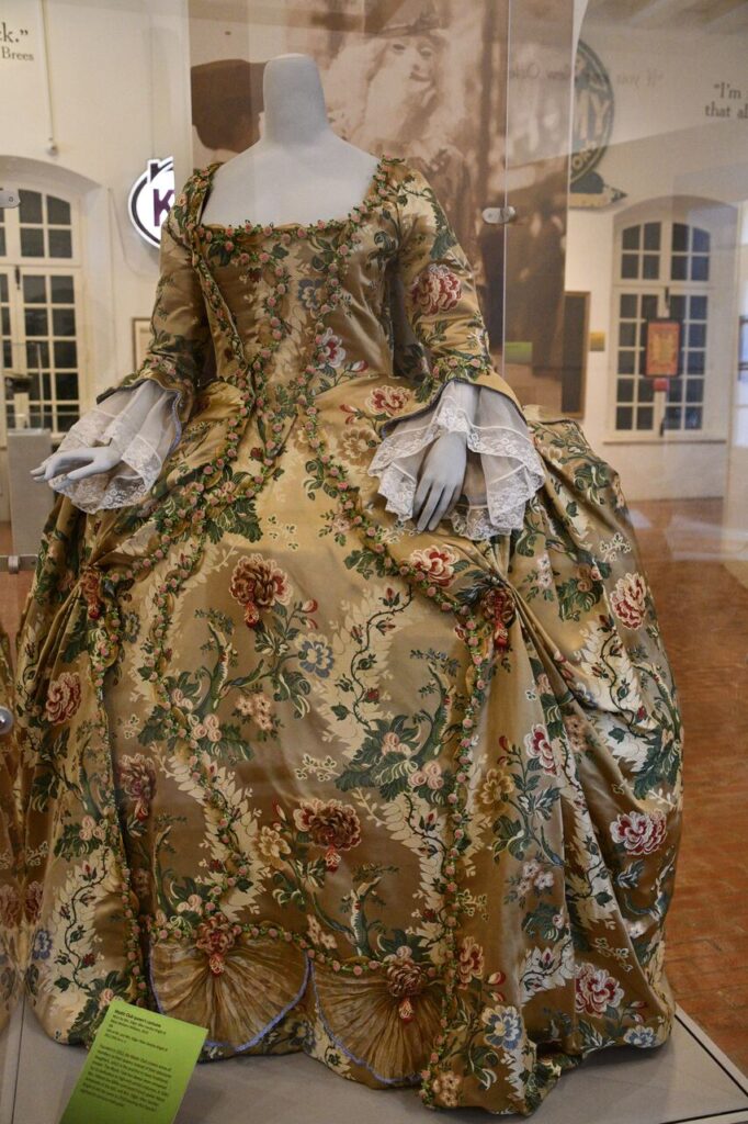
The 18th century. In the Louisiana State Museum in New Orleans, inside the Cabildo building, a golden brocade dress stands on a mannequin. Heavy, stiff, covered in large flowers. What if this dress encodes the protein from which the armour of one of the world’s most common viruses is built?”
PART 1: THE DRESS
The 18th century. In the Louisiana State Museum in New Orleans, inside the Cabildo building, a golden brocade dress stands on a mannequin. Heavy, stiff, covered in large flowers. What if this dress encodes the protein from which the armour of one of the world’s most common viruses is built?
Image description: NEW ORLEANS, LA – NOV 22: The Cabildo, part of the Louisiana State Museum, in New Orleans, as seen on Nov 22, 2023. Ritu Manoj Jethani – Shutterstock.
On a mannequin in a museum case stands a dress made of heavy, golden brocade. The fabric is stiff — it does not drape softly, but holds its shape. Across its surface spread large flowers: roses, peonies, leaves — densely embroidered, raised, dominant.
The bodice is fitted, narrowing at the waist. The sleeves end at the wrists with lace cuffs (engageantes). The skirt expands widely to the sides.
What sets this dress apart is the heaviness and stiffness of the fabric — brocade holds its shape like architecture, not like clothing. And the floral pattern, which is not mere decoration. On the fabric we see large flowers — roses, peonies — but also many smaller flowers arranged in chains running from bottom to top, connected by stems and leaves. It is an entire system: large flowers, small flowers, stems linking them into continuous chains. Too precise an arrangement to be merely ornamental.
PART 2: THE PROTEIN
What Is HPV?
HPV (Human Papillomavirus) is one of the most common viruses in the world. Nearly every one of us will encounter it during our lifetime. There are over 200 types of this virus. Some cause common warts on the hands. Others can lead to serious diseases. HPV6 — the type discussed in this post — causes warts but does not lead to cancer.
The virus enters the body through tiny breaks in the skin. Its target is the cells on the body’s surface. The virus gets inside them and forces them to produce its own proteins — using the machinery of our cells like a factory.
What Does the Virus Look Like from the Outside?
Every virus needs armour — a shell that protects its genetic material. In HPV, this armour is built by a protein called L1. Five copies of this protein join together like five blocks, forming a five-sided unit called a pentamer. 72 such blocks assemble together into a complete sphere — the shell of the virus.
The L1 pentamer is the brick from which the entire viral armour is built. All 200+ types of HPV share the same overall pentamer shape — the same armour architecture. They differ, however, in the loops that protrude from the surface. These small differences determine where the virus attacks and what disease it causes.
What Does an Antibody Do?
When our immune system spots a virus, it produces antibodies — proteins that grab the virus by its armour and neutralise it. In the structure I analyse in this post (PDB: 8YEF), we see an antibody fragment (abbreviated: Fab) attached to the L1 pentamer. This is the moment of defence — our body has grabbed the virus by its armour.
How Is the Pentamer Built?
The structure of this protein is publicly available in the protein database under the entry PDB: 8YEF. Anyone can view it in the free program Mol* at molstar.org.
source: RCSB Protein Data Bank, 8YEF. Visualisation: Mol* (molstar.org).
The antibody fragment Fab sits on top of the pentamer, attached to one of the surface loops. Fab consists of two chains — heavy and light — interlocked into a single compact structure.
The L1 pentamer consists of five identical protein chains arranged in five-fold symmetry. Each chain has a characteristic fold called „jelly-roll” — built mainly from beta sheets. Beta sheets are flat, rigid structures (in contrast to the spiral, flexible alpha helices). From the pentamer surface, loops protrude — variable regions that differ between HPV types and determine which antibodies can attach.
Below you will find a 3D animation showing how the HPV virus infects skin cells and how warts form:
[VIDEO: What are warts (HPV)? — 3D animation, AmerraMedical, YouTube]
PART 3: THE DECODING
Level 1: Protein Diagram (Front View)
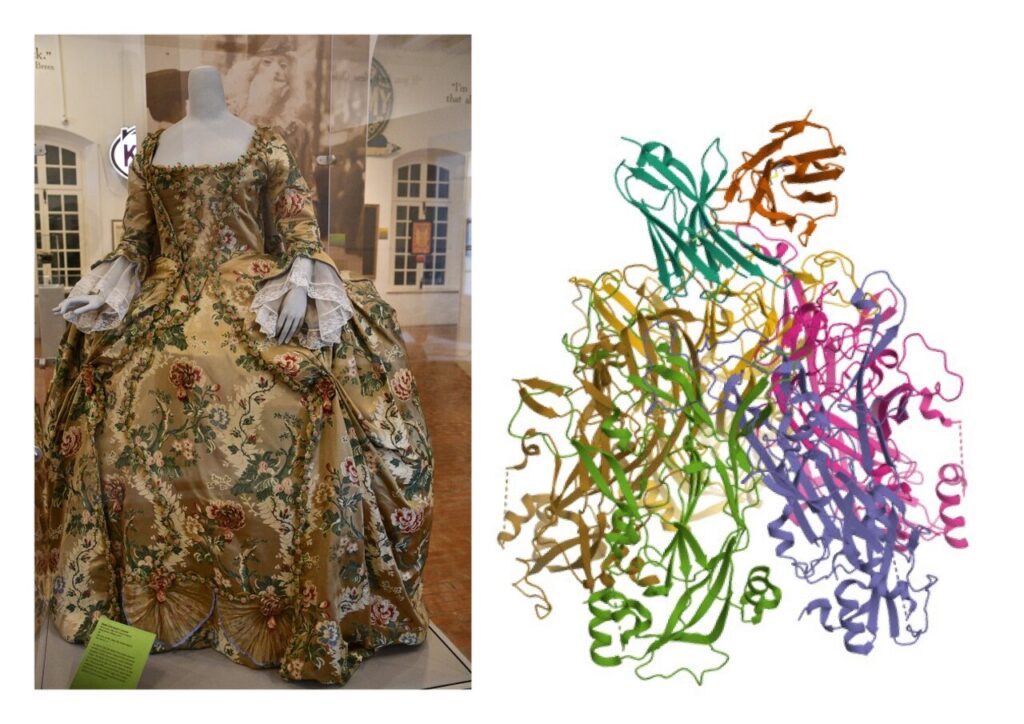
In protein visualisation software, a protein can be displayed as colourful ribbons and coils — a simplified diagram showing how the protein is built in space. Scientists call this the „cartoon representation.”
source: Side-by-side comparison — Cabildo dress (New Orleans) and protein 8YEF, ribbon view.
What corresponds to what?
Golden fabric background = protein surface The smooth, golden surface of the brocade is the surface of the pentamer — the base on which all structural elements of the protein are mounted.
All floral elements = protein structures visible on the surface On the golden background we see an entire decorative system: large flowers (roses, peonies), smaller flowers arranged in chains running from bottom to top, stems and leaves linking them into continuous paths. This is not decoration. It is a map of the protein’s structure. The large flowers correspond to the large loops protruding from the pentamer surface — the very ones that antibodies grab hold of. The smaller flowers arranged in chains correspond to polypeptide chains — continuous paths of amino acids running through the protein. The stems connecting the flowers correspond to linkers between structural elements. The entire floral pattern on the dress is a map of alpha helices, beta sheets, loops and chains — all the elements from which the protein is built.
Interestingly, all 200+ types of HPV share the same pentamer shape, but differ precisely in these surface elements — like dresses of the same cut, but with different flowers, patterns and decorations on the fabric.
Bodice made of two halves = antibody (Fab) The bodice is made of a left and right half sewn together. The Fab antibody is built the same way — from two chains (heavy and light) interlocked into a single whole. The bodice holds and squeezes — the Fab holds and squeezes the pentamer.
Lace cuffs (engageantes) = peripheral elements of Fab The cuffs on the sleeves are a separate element attached to the main structure. On the Fab protein, protruding elements are also visible at the periphery — fine structures at the edges of the antibody.
It is worth noting that historical dresses had many types of sleeves — long, short, lace-trimmed, or none at all. Our immune system also produces different classes of antibodies, each with a slightly different structure and function. Different sleeves may encode different types of antibodies — a topic for further investigation in future parts of „The Wardrobe of the Genome.”
Skirt expanding to the sides = wide base of the pentamer The pentamer seen from the front has a wide base. The dress skirt expands in exactly the same way.
Level 2: Spacefill Representation — Side View
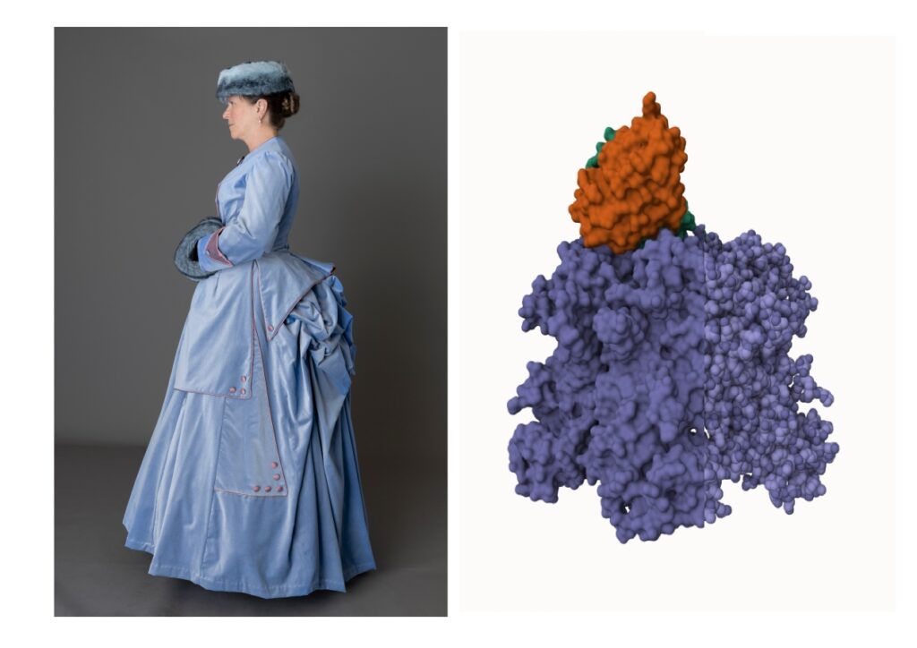
In Level 1 we saw the protein as ribbons and coils — a simplified diagram showing chains and helices. Now we are looking at the same protein, but in a different way. The „spacefill” representation shows every atom of the protein as a sphere at its real size. We no longer see chains or helices — we see the full volume that the protein actually occupies in space. This is the closest to what the protein „really” looks like — a dense, compact body of matter built from thousands of atoms of carbon, nitrogen, oxygen, hydrogen and sulphur. Scientists call this the „spacefill and surface representation.”
The Cabildo dress shows the protein from the front. But a protein also has a side profile — and to show it, a dress seen from the side, on a living figure, is needed. That is why in this comparison I used a different historical dress — worn by a woman, photographed in profile — which reveals the lateral shape and a key element: the protrusion at the back.
source: Side-by-side comparison — dress seen in profile and HPV6 L1 pentamer with Fab (PDB: 8YEF), spacefill representation, side view.
Image description: A Victorian woman wearing a pale blue 1870s bustle ensemble with a fur hat and muff. KathySG – Shutterstock.
In the spacefill side view, the protein reveals two distinct masses: a large purple body (the L1 pentamer) and a smaller orange body (the antibody fragment Fab). What is crucial is the position of the Fab — it does not sit on top of the pentamer centrally, but at its edge, on the periphery of the structure.
The dress seen in profile shows an identical arrangement. The bodice (Fab) does not dominate the skirt centrally — it is positioned at the edge of the skirt mass (the pentamer), shifted toward the periphery. Exactly as the Fab attaches to a surface loop on the rim of the pentamer, not at its centre.
But the key discovery is at the back of the dress.
In the protein image we see a deliberately split view. The left side of the protein is shown as a smooth, continuous surface (the „surface” view) — it corresponds to the front and sides of the dress, where the fabric is spread evenly and reveals the smooth external shape. The right side of the protein is shown as atomic filling (the „spacefill” view) — it corresponds to the back of the dress, where the fabric gathers, folds and pushes outward, revealing the internal architecture.
The front of the dress says: „this is what the protein looks like from the outside.” The back of the dress says: „this is how the protein is folded on the inside.”
Now it becomes clear why the ancients created these protrusions at the back of their dresses. The bustle, the draping, the padding — all these elements that fashion historians treat as aesthetic whims reveal their true function: they recreate the side profile of the pentamer structure and show the protein’s folding.
Anyone who has ever watched costume dramas — westerns, historical films, novel adaptations — has surely asked themselves the question: why are these dresses so wide? Especially at the back? Surely moving around in something like that every day must cause enormous difficulty. Generations of viewers have asked themselves this question. And the answer is simple: these dresses were not designed for comfort. They were designed to recreate the structure of a protein. The width, the protrusion, the folds — all of it has its molecular justification.
The 8YEF structure is a complex of two proteins: the L1 pentamer (a viral protein) and Fab (a human antibody). In the dress we see the same arrangement — the bodice (Fab) sits on top of the skirt (the L1 pentamer), attached to it at a specific point. Two separate elements joined into a single whole — just as two proteins are joined into a single complex.
This is the same profile. The same proportion of masses. The same logic of construction.
View from Below — the Interior of the Pentamer
A dress of the same type, suspended from the ceiling, reveals what is normally hidden — densely folded fabric visible both on the outside and underneath the skirt. The folds of fabric form a smooth but dense surface — one layer upon another.
photo description:New York, NY – December 4, 2017: Quinceanera dress by Mitzy on display at HBO screening and presentation of 15: A QUINCEANERA STORY at The Garage
source: Side-by-side comparison — dress suspended, view from below the skirt, and HPV6 L1 pentamer (PDB: 8YEF) tilted in Mol*, revealing the interior of the structure.
In Mol*, I tilted the 8YEF pentamer structure and switched to the „surface” view to look inside it from below. The result is identical: a smoothly folded, dense molecular surface filling the interior of the pentamer. We no longer see chains, helices or individual atoms — we see a continuous, folded molecular surface. Exactly like the folded fabric of the skirt.
The dress and the pentamer reveal the same thing: a folded surface visible from every angle — from the outside and from within. The dress from the front shows the protein’s surface. The dress from the side shows its profile. The dress from below shows the interior — the same folded structure seen from underneath.
Why Three Dresses?
In this post I used three separate dresses to show the protein from every angle. The dress from the Cabildo in New Orleans shows the protein from the front — it reveals the surface, the loops and the structure of the armour. The dress seen in profile shows the lateral shape and reveals the folding at the back. The suspended dress shows the interior — the dense, folded surface hidden underneath.
Finding a single dress photographed from the front, from the side and from below is extremely difficult. But there is also a deeper reason: each of these dresses has a different type of sleeves, a different length, a different bodice — and these differences may encode variants of the protein or different classes of antibodies. Three dresses are not a limitation. They are three views and potentially three variants of the same molecular complex.
CONCLUSIONS
The 18th-century dress from the Cabildo in New Orleans recreates the structure of the L1 pentamer — a building block of the HPV6 viral armour — in complex with the antibody fragment Fab (PDB: 8YEF).
The golden brocade corresponds to the rigid structures of the pentamer. The large flowers correspond to the surface loops. The bodice made of two halves corresponds to the antibody made of two chains. The side profile of the dress with its protrusion at the back mirrors the profile of the protein. The view from below reveals the folded surface of the interior.
But why does a viral protein belong in „The Wardrobe of the Genome”? Because the genome sews all garments — even those that are not its own design. The HPV virus cannot produce anything on its own. To create the L1 protein, it must enter a human cell and insert its DNA into the cell’s genome. From that moment on, it is our cell — our machinery — that produces the viral protein. The genome is forced to sew someone else’s garment.
This broadens the meaning of the entire series. In the first part, Elizabeth I’s dress encoded cholera toxin — a protein produced by the genome of a bacterium. In this part, the dress from New Orleans encodes the L1 pentamer — a viral protein produced by the human genome after the virus has hijacked it. „The Wardrobe of the Genome” covers all proteins — „ours” and „imposed.” The genome sews them all. Even those that are not its own design.
The structure of the L1 pentamer was solved in the 21st century.
The dress has been waiting since the 18th century.
Image search and matching, analysis and elaboration: Tomasz Mikulski – Cell God: 07/2026