„In the first part of „The Wardrobe of the Genome,” Elizabeth I’s dress on the Ditchley Portrait encoded cholera toxin (PDB: 1XTC). In the second, a golden gown from The Cabildo concealed the HPV pentamer (PDB: 8YEF).”
Now it is not a dress. It is a hairstyle.
In the title image you see a page from an eighteenth-century Parisian fashion catalogue — the „Galerie des Modes et Costumes Français” — with four portraits of women in elaborate hairstyles. The figures on these plates do not depict specific people — they are anonymous models illustrating the latest styles, drawn „d’après nature” (from life). But the creations they wear are the work of Rose Bertin and the circle of designers associated with the court of Marie Antoinette. I surrounded this page with living flowers, because you are about to see that the flowers on those hairstyles are not decoration. They are molecules.
In this entry I decode one of those four portraits: „Pouf à l’Asiatique” — upper right. The remaining three await their turn.
Eighteenth-century France. Paris. An era in which a woman wore on her head a tower of hair, feathers, flowers, and pearls — a construction so complex that it required an architect, not a hairdresser.
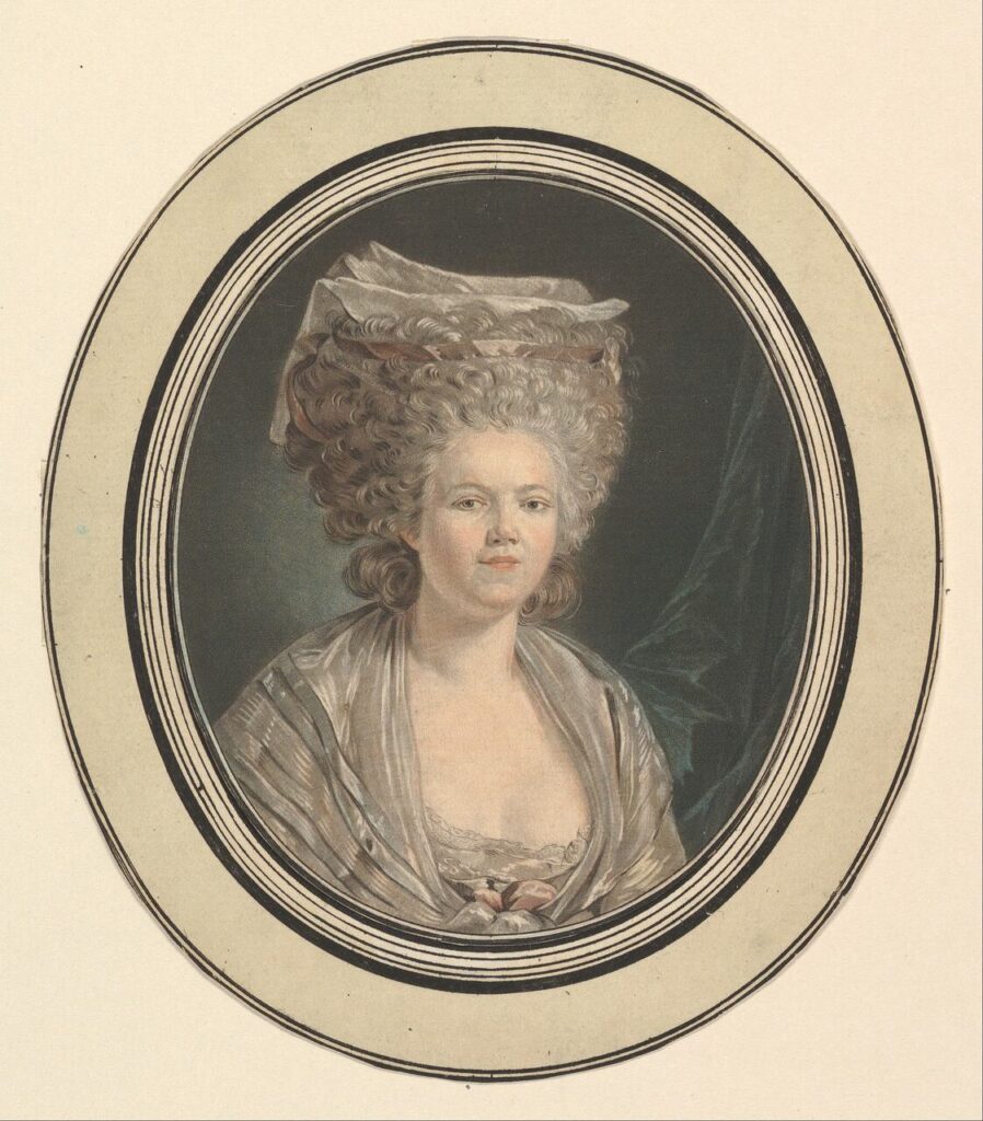
That architect truly existed. Her name was Marie-Jeanne Bertin — known to the world as Rose Bertin, although she never bore that name in her lifetime. „Rose” was given to her by nineteenth-century historians in apocryphal memoirs from 1824.
source: Mademoiselle Rose Bertin, Dressmaker to Marie-Antoinette – The Metropolitan Museum of Art.
She was born on 2 July 1747 in Abbeville and died on 22 September 1813 in Épinay-sur-Seine. She was the first recognised fashion designer in European history — the „Ministre des Modes” of Marie Antoinette. Twice a week she met with Marie Antoinette in private sessions, without ladies-in-waiting, designing creations that dictated fashion from Paris to Saint Petersburg.
The protein she encoded in a hairstyle is responsible for your every thought. Every memory. Every act of learning. It is the AMPA receptor — the synaptic gateway of the brain.
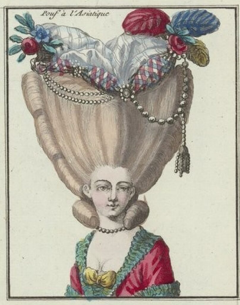
1. The Portrait — „Pouf à l’Asiatique”
[IMAGE: engraving „Pouf à l’Asiatique” from Cahier des Costumes Français, full]
The engraving comes from the „Cahier des Costumes Français” — a fashion catalogue published by Esnauts et Rapilly on rue St-Jacques in Paris. Source: Bibliothèque nationale de France (Gallica), public domain.
The woman in the portrait wears a bodice in turquoise and pink, strings of pearls around her neck and between the layers of her hairstyle, and at the top of her head — a decoration called a „pouf”: a towering construction of hair, fabric, feathers, flowers, and ribbons. The inscription at the top reads: „Pouf à l’Asiatique.”
At the bust, a yellow bow is visible — tied and cinched in the middle, as if closing or squeezing something shut.
From the vicinity of the hairstyle hangs a tassel (passementerie) — an ornamental cord ending in fringes, falling alongside the hair.
On the edges of the pouf, flowers with green leaves are visible.
2. The Protein — AMPA Receptor GluA2 (PDB: 6RUQ)
[IMAGE: 6RUQ in Mol*, cartoon view, four chain colours]
The AMPA receptor (α-amino-3-hydroxy-5-methyl-4-isoxazolepropionic acid) is a ligand-gated ion channel — one of the most important proteins in the central nervous system. It is responsible for the majority of fast excitatory synaptic transmission in the brain: when a neuron releases glutamate (the most important excitatory neurotransmitter), the AMPA receptor in the postsynaptic neuron opens and admits ions, generating an electrical signal. Without this protein there is no thought, no memory, no learning.
The name „AMPA” comes from a laboratory substance that can artificially open this gate — but in the living brain the key is always glutamate, the most important excitatory neurotransmitter.
The protein has a tower-like structure — three storeys stacked one above the other, from bottom to top:
TMD (transmembrane domain) — the „basement” embedded in the cell membrane. This is where the channel lies — the tunnel through which ions flow. Four subunits of the protein surround a central pore like four columns around a shaft.
LBD (ligand-binding domain) — the „main floor,” where glutamate binds to the protein. This is where the decision is made: if glutamate arrives and „sits” in the right place, the protein changes shape and opens the channel below.
ATD (amino-terminal domain) — the „tower” at the very top. The largest and most elaborate part of the structure. It is responsible for assembling the receptor from four subunits and for regulating its behaviour.
The AMPA receptor belongs to the family of ionotropic glutamate receptors (iGluR) — one of the most important protein families in the brain. This family is divided into four subfamilies: AMPA, kainate, NMDA, and delta. All share the same three-layered architecture (TMD–LBD–ATD) but differ in their details — like four portraits on a single page of a fashion catalogue, each with the same silhouette but different decorations.
Structure 6RUQ is a GluA2 receptor from the rat brain, captured in a sleeping state — blocked by two substances that prevent it from opening. It is like a photograph of a closed gate.
But in the living brain this gate opens millions of times per second:
[IMAGE: AMPA receptor during activity — glutamate (orange) activates the receptor, ions (red spheres) flow through the channel into the neuron. Source: PDB 3KG2, 3D rendering / Shutterstock]
In this graphic the receptor is shown in action: glutamate molecules (orange) sit in the LBD domain, the gate is open, and red spheres — ions — flow through the TMD channel into the cell. This is the moment a nerve signal is born.
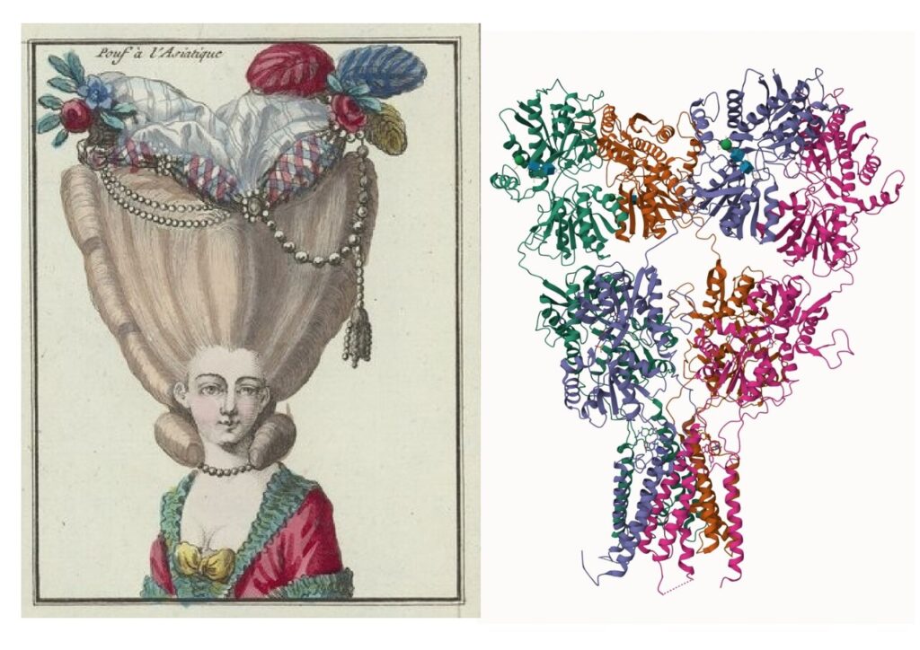
3. Decoding — Level 1: Silhouette (cartoon)
[IMAGE: side-by-side — Pouf à l’Asiatique engraving vs 6RUQ cartoon]
The silhouette of the protein and the silhouette of the portrait show a striking similarity across three layers:
ATD — amino-terminal domain, crown of the structure
Curled hair at the neck and sides of the hairstyle
Alpha helices — spiral secondary structures of the protein
Strings of pearls hanging between layers
Interdomain linkers (ATD–LBD and LBD–TMD)
Yellow bow at the bust — tied, cinched
Ion channel gate in the TMD — closed in the 6RUQ structure
Tassel (passementerie) hanging from the hairstyle
Glutamate — the ligand approaching the LBD domain
Hair as alpha helices
Near the neck and on the sides of the hairstyle, the hair is arranged in characteristic curls — looped, spiral coils. In the cartoon diagram alongside, exactly the same motif is visible: alpha helices — spiral structures into which segments of the polypeptide chain coil. This is the fundamental building block of the protein. In the cartoon view, the AMPA receptor is full of these spirals — and the „Pouf à l’Asiatique” hairstyle reproduces them in hair.
[IMAGE: 6RUQ molecular surface, front view — orange-red glycans on the green ATD surface]
When the view in Mol* is switched from „cartoon” to „molecular surface,” the ribbons and helices vanish, replaced by the smooth surface of the protein. And on that surface, in the ATD zone, distinct orange-red patches are visible, scattered across the edges of the upper part of the protein.
These are glycans — short sugar chains that the cell attaches to the protein after it has been produced. They serve as „calling cards” — helping other molecules recognise the protein, protecting it, and sometimes regulating its function. On the molecular surface they appear as small protuberances scattered across the edges of the structure — like flowers on the hairstyle decoration.
Now look at the pouf:
On the edges of the hairstyle decoration, two red flowers with green leaves are visible — positioned at the outer edges, not at the centre. Exactly as the glycans sit on the exposed surface of the ATD — at the edges, not at the centre. Two flowers, two glycans visible from the front.
Rose Bertin encoded not only the general pattern of glycosylation — not only the fact that the protein „wears flowers” — but even their number and placement on the edges of the crown.
The tassel — glutamate
From the vicinity of the hairstyle, at the boundary between the ATD and LBD layers, an ornamental tassel (passementerie) hangs. In the portrait it is attached to one of the strings of pearls — it does not hang freely but is connected to the chain. This is not accidental: in the protein, glutamate binds in the pocket of the LBD domain, which is structurally connected to the linkers (strings of pearls). The tassel hangs at the binding site — attached to the linker, precisely where the ligand connects with the protein.
In the living brain, glutamate — a small molecule released by the sending neuron — reaches the receptor and „enters” the LBD pocket, triggering the opening of the channel. The tassel in the portrait encodes this molecule: it hangs from the pearl chain in the LBD zone, ready to act.
The yellow bow — the channel gate
At the bust, at the boundary of the TMD layer, a yellow bow is visible — tied and cinched in the middle. This gesture of tying is not ornament. The AMPA receptor in the 6RUQ structure is blocked — the ion channel is closed, no ions can pass through. The bow tied in the middle symbolises this state: the gate cinched shut, the channel closed.
When glutamate (the tassel) reaches the LBD domain and binds to the protein, the bow „unties” — the channel opens and ions flow into the neuron. This moment — the transition from closed to open gate — is visible in the active receptor graphic in section 2 above: red ions flow through the channel, glutamate sits in the LBD pocket, the bow is „untied.”
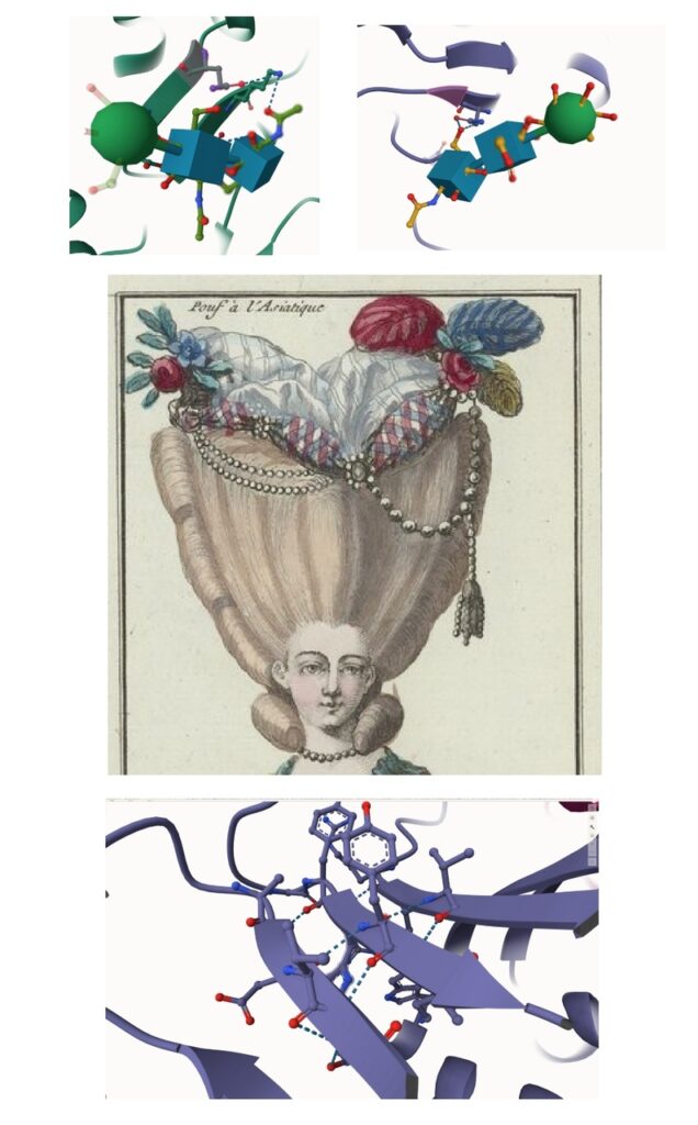
5. Decoding — Level 3: Close-up
[IMAGE: close-up of glycans in Mol* — SNFG representation: green spheres and turquoise cubes]
In magnification, the glycans appear in SNFG representation (Symbol Nomenclature for Glycans) — the standard visual code used in Mol*:
Green spheres = mannose (BMA) — corresponding to the flowers on the pouf: rounded, full, like petals
Turquoise/blue cubes = N-acetylglucosamine (NAG) — corresponding to the branch points in the flower stems: angular, geometric points where the sugar chain forks
In the portrait, the flower stems are not visible — they are hidden beneath the pouf decoration, under the fabrics and ribbons. But in the molecular structure we see them clearly: the NAGs form the „trunk” of the glycan, from which mannose grows like a flower at the end of a stem.
[IMAGE: close-up of protein structure in Mol* — beta sheets, aromatic rings, hydrogen bonds]
The second close-up reveals the internal structure of the protein in the zone between domains. Three elements encoded in the portrait are visible here:
Beta sheets — flat, ribbon-like structures arranged in parallel, like layers of fabric. In the portrait they correspond to the fabrics above the hair — the pouf material with its characteristic pattern of overlapping rhombuses. These rhombuses are not ornament: in the protein, helices and beta sheets form exactly such a lattice — regular, repeating secondary structures woven into a geometric pattern.
Aromatic rings — flat, hexagonal structures of aromatic amino acids (phenylalanine, tyrosine, tryptophan), visible in magnification as hexagons surrounded by smaller spheres. In the portrait this is the brooch — a central ring-shaped element surrounded by small circles. The circles are not electrons — they are atoms surrounding the ring, exactly as in the portrait of Elizabeth I, where gold medallions encoded atoms on the protein surface and the necklace encoded an aromatic ring. The same code, a different era. Aromatic amino acids form the hydrophobic core of the protein, stabilising the entire structure through π–π interactions (ring stacking) — they are what „holds together” the domain.
Amino acid chains with polypeptide backbone — running regularly, with red oxygen atoms and blue nitrogen atoms protruding sideways, connected by blue dashed lines of hydrogen bonds. In the portrait these are the strings of pearls— chain-like, regular, with small elements branching sideways like tassels.
The pearls in the portrait appear in three different forms:
First chain — a thicker string of pearls around the neck, encircling the zone of transition between the face (LBD) and the chest (TMD). These are the LBD–TMD linkers (S1-M1, M2-S2, S2-M3), connecting the ligand domain to the ion channel. The same type of chain appears on the right side of the brooch — and it is from this chain that the tassel (glutamate) hangs, attached to the linker at the binding site.
Second chain — a string of pearls hanging between the pouf and the hair. This is the ATD–LBD linker, connecting the crown to the middle storey.
Third chain — finer and double, found on the opposite side of the brooch from the tassel. Two thin strings running in parallel. In the protein this corresponds to hydrogen bonds between beta sheets — visible in the close-up as blue dashed lines connecting two parallel polypeptide strands. The double pearl chain encodes the dual nature of the beta sheet: two strands running side by side, fastened by regular hydrogen bonds, thinner and denser than the main polypeptide backbone.
Three feathers at the top of the pouf? 🤔
At the very apex of the decoration — above the flowers and fabrics — three feathers in different colours protrude: red/crimson, blue, and olive-yellow (a leaf). They sit at the summit of the ATD.
What do they encode? I do not yet know.
The number three may refer to the three main structural elements of the receptor — three layers (TMD, LBD, ATD) — symbolically encoded at the summit, like an emblem summarising the entire edifice. But this is a hypothesis, not a certainty. Perhaps the answer will come from subsequent portraits in the same catalogue — if other hairstyles encode other proteins from the same family, comparison between them will establish whether the three feathers are a constant shared element or a variable detail specific to this particular protein.
6. Conclusions — how many proteins does the wardrobe hold?
In the „Wardrobe of the Genome” series we discover proteins hidden in hairstyles and garments. But on CellGod.live I have already decoded over a dozen proteins — and not only in fabrics. Proteins have been encoded in sculptures, in human and animal figures, in tools, in heraldic symbolism, in military insignia, in religious artefacts. Clothing is only one carrier. The genome, by human hand, inscribed its molecules everywhere — in stone, wood, metal, on canvas, and in architecture.
How many proteins does the genome produce in total? How many molecules await discovery in the works of humanity?
The human genome contains approximately 20,000 protein-coding genes — 20,000 „recipes” for the molecules from which the body is built. The international Human Proteome Project (HPP) spent over a decade searching for experimental evidence for the existence of each of these proteins. In 2020 the HPP consortium announced in Nature that it had confirmed the existence of proteins for 17,874 genes — approximately 90% of the entire set. The remainder are „missing proteins,” probably so rare that they occur only in specific tissues or within a narrow developmental time window.
But 20,000 is only the first level.
In reality, one gene does not equal one protein. The genome can create many versions of the same protein from a single recipe — so-called proteoforms. How? In three ways.
First, through alternative splicing — the same gene can be „read” in different ways, skipping certain segments, yielding from several to a dozen or more variants. This raises the number to 70,000–100,000 different protein sequences.
Second, through post-translational modifications — after producing a protein, the cell attaches „add-ons” to it: phosphate groups, sugar chains (glycosylation — exactly what we saw on the pouf as flowers!), ubiquitin tags, and dozens of others. Each combination yields a functionally different molecule. A single amino acid chain can exist in hundreds of differently modified versions.
Third, through genetic polymorphisms — natural differences in DNA between individuals. Your AMPA receptor and my AMPA receptor may differ slightly, because our genomes are not identical.
The sum of these three mechanisms yields estimates of 250,000 to as many as one million proteoforms in the human body. The „Human Proteoform Project” aims to catalogue them in full.
The genome did not encode 20,000 proteins. It encoded a million. And it hid them not only in garments — but in the entire material culture of humanity.
And this is precisely what we see in this entry. The flowers on the pouf are not decoration — they are glycosylation, one of the post-translational modifications that turns one protein into many versions of the same protein. Flowers on an eighteenth-century hairstyle are two sugar chains on the surface of the receptor — visible to the naked eye on an eighteenth-century engraving.
The genome sews all the garments. And Marie-Jeanne Bertin knew perfectly well what proteins look like — two and a half centuries before science gave them that name.
Image search and matching, analysis and elaboration: Tomasz Mikulski – Cell God: 07/2026
BIBLIOGRAPHY Molecular structures and biology Berman, H. M., Westbrook, J., Feng, Z., Gilliland, G., Bhat, T. N., Weissig, H., … & Bourne, P. E. (2000). „The Protein Data Bank.” Nucleic Acids Research, 28(1), 235–242. https://www.rcsb.org/ Protein Data Bank. (2024). „6RUQ: Structure of GluA2cryst in complex with the antagonist ZK200775 and the negative allosteric modulator GYKI53655 at 4.65 Å resolution.” https://www.rcsb.org/structure/6RUQ Krintel, C., Dorosz, J., Larsen, A. H., Thorsen, T. S., Venskutonyte, R., Mirza, O., Gajhede, M., Boesen, T., Kastrup, J. S. (2021). „Binding of a negative allosteric modulator and competitive antagonist can occur simultaneously at the ionotropic glutamate receptor GluA2.” FEBS Journal, 288, 995–1007. Traynelis, S. F., Wollmuth, L. P., McBain, C. J., et al. (2010). „Glutamate receptor ion channels: structure, regulation, and function.” Pharmacological Reviews, 62(3), 405–496. Sobolevsky, A. I., Rosconi, M. P., & Gouaux, E. (2009). „X-ray structure, symmetry and mechanism of an AMPA-subtype glutamate receptor.” Nature, 462, 745–756. Proteome and proteoforms Kim, M. S., Pinto, S. M., Getnet, D., et al. (2014). „A draft map of the human proteome.” Nature, 509, 575–581. Omenn, G. S., Lane, L., Overall, C. M., et al. (2020). „Progress Identifying and Analyzing the Human Proteome: 2020 Metrics from the HUPO Human Proteome Project.” Journal of Proteome Research, 19, 4735–4746. Aebersold, R., Agar, J. N., Amster, I. J., et al. (2018). „How many human proteoforms are there?” Nature Chemical Biology, 14, 206–214. Ponomarenko, E. A., Poverennaya, E. V., Ilgisonis, E. V., et al. (2016). „The Size of the Human Proteome: The Width and Depth.” International Journal of Analytical Chemistry, 2016, 7436849. Fashion history Bertin, M.-J. (1824, apocryphal). Mémoires de Mlle Bertin sur la Reine Marie-Antoinette. Attributed to Jacques Peuchet. Weber, C. (2006). Queen of Fashion: What Marie Antoinette Wore to the Revolution. Henry Holt and Company, New York. Ribeiro, A. (2002). Dress in Eighteenth-Century Europe, 1715–1789. Yale University Press. Esnauts et Rapilly (pub.). Cahier des Costumes Français. Paris, rue St-Jacques. Source of engravings: Bibliothèque nationale de France, Gallica (public domain).
Mol* (molstar.org). „Mol*: Molecular Structure Viewer.” PDBj. Tool for protein structure visualisation used in the analysis of 6RUQ. UniProt Consortium. „GluA2 / GRIA2 — Glutamate Receptor Ionotropic, AMPA Type Subunit 2.” https://www.uniprot.org/uniprot/P19491