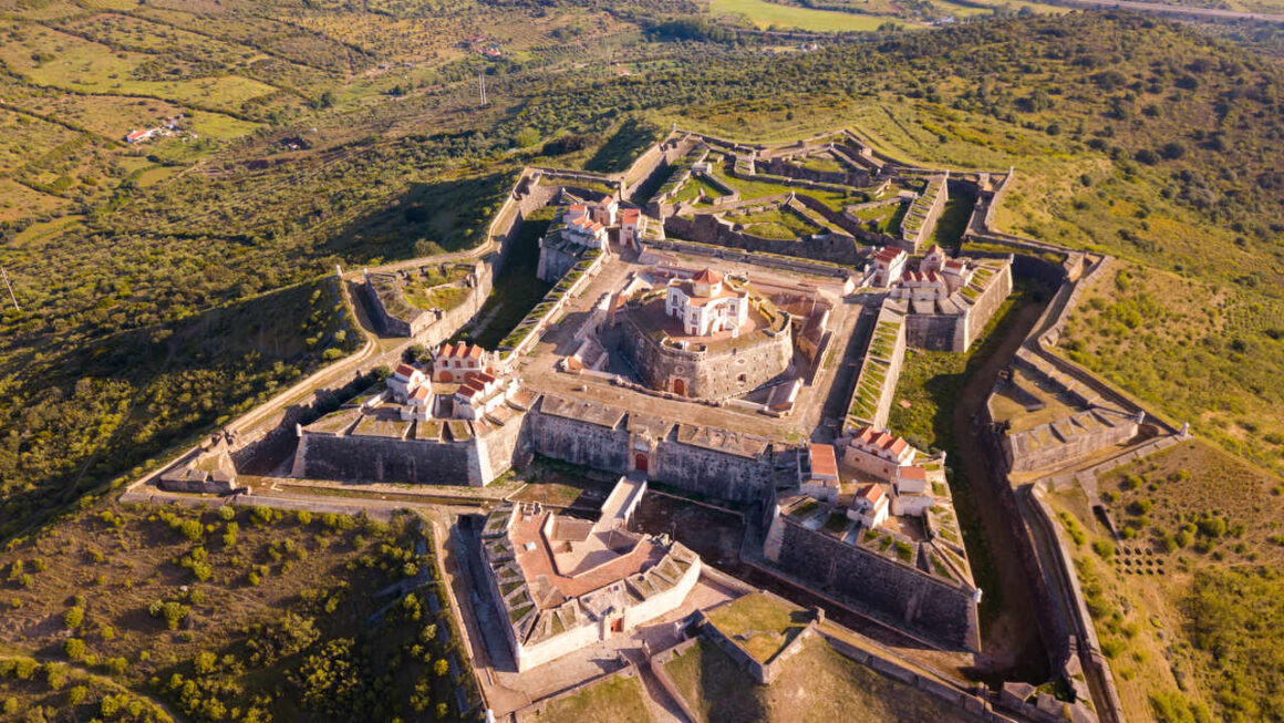
Fort Nossa Senhora da Graça in Portugal is a bastioned fortification built between 1763 and 1792. Officially called a „masterpiece of 18th-century military architecture,” it rises on Monte da Graça hill (404 m above sea level) near the city of Elvas.
Technical parameters:
Star-shaped plan with fortifications and bastions
Three concentric defensive lines
Never conquered by an enemy
UNESCO World Heritage Site (since 2012)
Fort = Microglia
Photo description: Aerial view of star-shaped fortification of Nossa Senhora da Graca Fort in village of Alcacova, Portalegre, Portugal. BearFotos – Shutterstock.
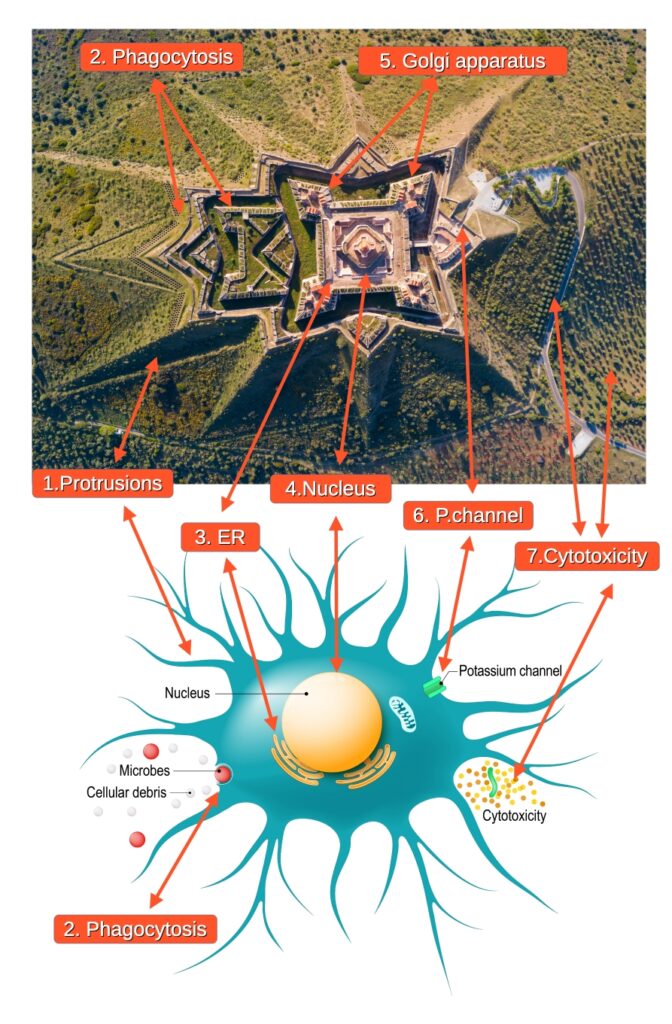
Vector description: Anatomy of a microglial cell. glial cell is the macrophage for immune defence the central nervous system. Vector diagram for educational, medical, biological and science use. Designua – Shutterstock.„
The bird’s-eye view reveals the fort’s identity with microglia – the brain’s macrophages. The star shape with radiating processes, multi-layered defensive structure, and central nucleus create an exact map of this immune cell.
Microglia constitute 10-15% of all brain cells. They form the first line of defense of the nervous system – phagocytosing pathogens, removing dead cells, and controlling inflammation.”
Functional map: From phagocytosis to exocytosis
Fort Nossa Senhora da Graça encodes POLARIZED microglia – a cell with a distinct front and rear, actively migrating and phagocytosing pathogens.
Ravelins, glacis, counterscarp – projecting outward from the fort
Branches extending radially
Recognition function and first line of defense
Dynamic scanning of terrain (1–2 μm/min – a single protrusion moves the thickness of one bacterium per minute, yet thanks to numerous protrusions the entire microglial cell scans its surroundings every 3–4 hours).
2. FRONT OF THE CELL (Leading Edge) → Phagocytosis
This is the mouth of the microglia. Here the cell makes contact with the enemy – „microbes, cellular debris” – i.e., pathogens and cellular remnants it must eliminate. From scanning protrusions, pseudopodia are formed – structures that envelop and draw the enemy inside.
Crownwork = MOUTH and PHAGOSOME This grand, three-branched fortification pushed farthest forward is the MOUTH of the microglia. Here enter „microbes” and „cellular debris” – everything the cell must engulf and destroy. Once the enemy is drawn inside, the Crownwork closes around it and becomes a PHAGOSOME – a sealed death chamber. The pathogen is not attacked externally. It is swallowed, sealed in this chamber, and only then destroyed. Simply: Crownwork = a mouth that swallows the enemy and locks it in a death chamber.
Hornwork INSIDE the Crownwork = Killing Machinery Inside the Crownwork lies the Hornwork – a smaller, two-horned structure. It is not a separate building, but the interior of the death chamber. The „two bull’s horns” are enzyme complexes embedded in the phagosome walls – these destroy the enemy in the isolated chamber. Among them is NOX2 (NADPH oxidase), an enzyme producing reactive oxygen species – molecular „projectiles” piercing the pathogen’s membrane.
3. Parapet of the Redoubt = Endoplasmic Reticulum (ER)
The square structure surrounding the nucleus is the endoplasmic reticulum (ER) – an extensive network of tubules and cisternae inside the cell. In biology, the ER is a direct extension of the outer nuclear envelope membrane. Here, on ribosomes, proteins are synthesised based on instructions from DNA (transcription → translation).
Ribosomes = Soldiers and Gunsmiths
The ER is a factory, but every factory needs workers. In biology, these are ribosomes – tiny protein-RNA complexes that read instructions from the nucleus (mRNA, the protein „recipe”) and assemble proteins accordingly. In the fort’s architecture, ribosomes are the soldiers and gunsmiths – people who physically produce ammunition (cytokines, enzymes, proteases) and prepare it for further processing in the Golgi Apparatus.
Ribosome (biology)
Soldier / Gunsmith (fort)
Reading mRNA (instructions from nucleus)
Reading command orders
Protein synthesis (ammunition)
Producing ammunition and weapons
Working on the ER surface
Working in warehouses and barracks
Passing proteins to the Golgi
Delivering weapons to the casemates
Without ribosomes, there is no ammunition. Without soldiers, there is no defence.
4. Nucleus → Central redoubt with dome
The command centre of the entire structure. Contains DNA – the complete blueprint for building and operating the cell.
Regulation of gene transcription – copying instructions from DNA to mRNA.
5. Fragmented Golgi Apparatus → Flanks with Casemates
This particular diagram of microglia omits the Golgi apparatus – it focuses solely on immune functions. But the fort reveals it.
The four corner flanks with their characteristic towering, spire-like roofs represent the fragmented Golgi Apparatus.
What are casemates? Architecturally, they are vaulted, shell-resistant chambers within the fort’s ramparts, serving as storage facilities and firing positions. Their towering, arched roofs correspond to the shape of Golgi cisternae – flattened, curved membranous sacs visible under an electron microscope.
In activated microglia, the Golgi apparatus is not a single compact structure. It undergoes fragmentation – breaking down into several smaller stacks of cisternae strategically dispersed throughout the cytoplasm. The fort exactly reflects this state: the four casemated flanks are four independent centres for modifying and packaging proteins (e.g., pro-inflammatory cytokines) into secretory vesicles.
Link to Exocytosis: It is within the Golgi Apparatus that proteins from the ER are sorted and packaged into vesicles, which then travel to the cell membrane. When a vesicle reaches the membrane, it fuses with it and expels its contents outside – this is exocytosis. The casemates (Golgi) are therefore where „ammunition” is prepared for firing. The actual „firing”, however, occurs not here, but on the ramparts – via the batteries.
Batteries on the Ramparts = Points of Exocytosis
On the external ramparts of the fort, along the entire perimeter, are positioned batteries – artillery emplacements directed outward. These are the points of exocytosis – sites through which the microglia ejects cytokines and chemokines (signalling proteins) to the outside, alerting other immune system cells.
This is a crucial observation: exocytosis does not occur solely at the rear of the cell, but along its entire perimeter – just as the batteries are distributed around the fort’s walls.
6. REAR OF THE CELL (Uropod) → Reduits + Posterns + Main Gate
The rear part of the microglia is not the main site of cytokine ejection, but rather a signalling centre – this is where the structures responsible for receiving the alarm signal and triggering the cascade of events leading to exocytosis are located.
Reduits = Calcium Stores
The fort contains numerous Reduits – „structures within a structure”, final points of resistance. One central Reduit is connected by a bridge to the Parapet (perinuclear ER). Further Reduits are distributed in the fort’s peripheral walls. All are interconnected, forming an extensive network – exactly like the Endoplasmic Reticulum (ER), which weaves throughout the entire cytoplasm.
In microglia, the Reduits are calcium stores (Ca²⁺) – intracellular reservoirs of calcium ions, the crucial alarm signal. The central Reduit by the Parapet is the perinuclear ER. The Reduits in the walls are the cortical (sub-plasma membrane) ER, which extends right beneath the cell surface. Ca²⁺ ions are among the most important signal transducers in the cell – their release launches a cascade of reactions leading to cell activation.
Posterns – Hidden Alarm Gates
The central Reduit is connected to the Parapet (ER) by a bridge containing two posterns – small, concealed gates, shut during peacetime. These are distinct, strategic passages linked to transport and communication between the perinuclear ER and the rest of the network.
Main Gate = Kv1.3 Potassium Channel
At the rear of the fort lies the Main Gate. In microglia, this is the Kv1.3 potassium channel – a specialised protein complex embedded in the cell membrane that enables a massive efflux of potassium ions (K⁺) to the outside.
When the calcium wave from the Reduits reaches the Main Gate, the Kv1.3 channel opens. The potassium efflux changes the electrical potential of the cell membrane (like flicking a switch) and triggers exocytosis – the batteries on the ramparts eject cytokines to the outside.
7. Cytotoxicity → Molecular Discharge
Destruction of enemies (pathogens, damaged cells).
Phagocytic function.
Vegetation around the fort = map of discharged ammunition distribution.
KEY DISCOVERY: Vegetation Around the Fort = Pattern of Molecular Discharge
Look at both aerial images:
MICROGLIA DIAGRAM:
Cytotoxicity visible as molecules released outward
Concentration of molecules in certain places (clusters)
Scattered molecules in other places (diffuse cloud)
FORT PLAN:
Vegetation around the fort (trees, bushes) in an identical arrangement
Dense tree clusters in some places = dense molecular concentration
Scattered trees elsewhere = diffuse molecules
EXACTLY THE SAME GEOMETRY:
Clusters vs. dispersion
The aerial view is identical
1:1 spatial distribution
The trees and bushes surrounding the fort constitute a map of the chemical molecule distribution released by microglia — concentrated in areas of intense attack and dispersed where activity is lower. Crucially, the vegetation is concentrated mainly at the rear of the fort (corresponding to the main secretory pathway ER → Golgi → Main Gate), but is also visibly spread all around – this is precisely the effect of the batteries, the points of exocytosis along the entire perimeter of the cell membrane.
Portugal 1763–1792: They even managed to immortalizethe dynamics of molecular discharge.
Military language in biology – not metaphor, but reality
Before we delve into the details of the fort’s weapons, we must understand something fundamental: modern immunology itself uses military terminology to describe immune cells.
Official scientific terms:
„Killer cells” (NK cells – Natural Killer)
„Cytotoxic attack”
„Molecular bullets”
„Chemical warfare” (ROS)
„Arsenal of cytokines”
„Immune soldiers”
„Killing chamber” (phagosome)
„Respiratory burst” = artillery barrage
Educational microscopy videos show:
Neutrophils „attacking” bacteria
Macrophages „devouring” the enemy
NK cells „firing” perforins like projectiles
Phagosomes as „execution chambers”
T cells „killing” infected targets
Fort Nossa Senhora da Graça does not use „metaphor” – it uses the literal language of defensive biology that 21st-century science itself recognizes as the most accurate description of immune cell function.
The Portuguese in the 18th century encoded the COMPLETE MICROGLIAL DEFENSE SYSTEM in fortress architecture – using the same terms (cannons, chambers, bombardment, arsenal) that modern immunology itself uses to describe molecular warfare.
This is not „similarity” – this is a MAP.
„The Interior of the Fort = The Microglia Nucleus
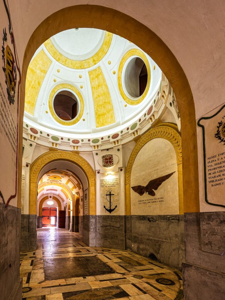
Photo description: Elvas, Portugal – Apr 02, 2025: Interior of the Fort Nossa Senhora da Graca or Fort Conde de Lippe north of the city of Elvas in Alentejo in Portugal.RediErnst – Shutterstock.”
Look at this interior of the fort. You are looking at the microglia nucleus. I’ve shown you this structure many times in previous posts — do you see the dome? That’s the nuclear envelope. Do you see those oval openings in the dome? Those are nuclear pores (Nuclear Pore Complex) — channels transporting proteins and RNA between the nucleus and cytoplasm.
But today we’ll focus on something else. On the eagle symbol beneath the dome. Because what the Portuguese placed in the fort’s central redoubt in 1939 is an encoded map of a key cell nucleus protein.
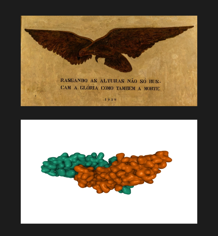
The Eagle Under the Dome = BMP-6
Beneath the dome, a majestic eagle with outstretched wings is visible. Below it, an inscription:
„RASGANDO AS ALTURAS NÃO SÓ BUSCAM A GLÓRIA COMO TAMBÉM A MORTE”
„Tearing through the heights, they seek not only glory but also death”
1939
Photo description: Elvas, Portugal – Apr 02, 2025: Interior of the Fort Nossa Senhora da Graca or Fort Conde de Lippe north of the city of Elvas in Alentejo in Portugal.RediErnst – Shutterstock.
Illustration description:Structure of bone morphogenetic protein 6 homodimer, 3D cartoon and Gaussian surface model, white background. VD Image Lab – Shutterstock.
Perfect anatomical matching
When you overlay the 3D structure of BMP-6 protein onto the eagle sculpture – everything matches exactly in the same place:
✓ Eagle’s head = Dimer center (cysteine knot) ✓ Left wing = Left monomer (cyan) ✓ Right wing = Right monomer (orange) ✓ Bird’s tail = Protein chain ends (N/C-terminals)
This is not a metaphor. This is a literal 3D map.
The Portuguese in 1939 carved BMP-6 protein with positional accuracy of every element – 60 years before its crystal structure was solved.
Compare:
| Eagle under the fort’s dome | ↔ | 3D structure of BMP-6 protein | | Two symmetrical wings | ↔ | Two identical protein subunits | | Extended form | ↔ | Active dimer conformation |
BMP-6 in the Microglial Nucleus — Scientific Confirmation
BMP-6 is found in microglial cells and translocates to the nucleus via the BMP/SMAD pathway:
Mechanism of Action:
BMP-6 binds to receptors on the microglial surface.
It activates the phosphorylation of SMAD1/5/8 proteins (pSMAD – phosphorylated, i.e., activated signalling proteins).
pSMAD + SMAD4 → translocation to the nucleus (movement of the signalling complex).
Inside the nucleus, this complex regulates gene transcription – switching concrete genetic programmes on or off.
Scientific Evidence:
Varga et al. (2021): „IL-6 regulates hepcidin expression via the BMP/SMAD pathway … in BV2 glial cells”
Immunohistochemical studies have shown „nuclear co-staining with pSMAD” in microglial cells.
BMP-6 is broadly present in the central nervous system.
BMP-6 is located in the microglial nucleus and regulates gene expression. The eagle beneath the dome (the nucleus) is literally a map of its location.
„Glory and Death” – The Dual Function of BMP-6
The Portuguese inscription literally describes this protein’s function in glial cells:
„GLÓRIA” (glory) = LIFE, REGENERATION:
Cell proliferation (multiplication)
Differentiation and development
Neural tissue regeneration
Neuroprotection following brain injury (protection of neurons from damage)
„BMP-6 expression is induced in astrocytes surrounding sites of brain injury, where it serves a neuroprotective function”
„MORTE” (death) = APOPTOSIS, ELIMINATION:
Programmed cell death (apoptosis)
Inhibition of proliferation
Elimination of damaged cells
Production of iNOS (inducible nitric oxide synthase – an enzyme producing nitric oxide NO, a gaseous messenger and chemical weapon) and TNF-alpha(tumour necrosis factor alpha – a potent pro-inflammatory cytokine) in macrophages, leading to B-cell death
BMP-6 decides whether the microglial cell will proliferate (regeneration, defence) or die (apoptosis, population control). It is a judicial protein, the arbiter of the cell’s fate.
„Tearing Through the Heights” — Transcriptional Activity
„RASGANDO AS ALTURAS” — tearing through the heights. This signifies:
Penetration to the Nucleus:
BMP-6 (via the SMAD pathway) reaches the „heights” = the cell nucleus.
It „pierces” the nuclear envelope through the nuclear pores.
It regulates transcription at the „highest point” of the cell.
Transcriptional Activity:
Inside the nucleus, pSMAD1/5/8 binds to DNA.
It „tears open” chromatin (the DNA-protein complex), opening genes.
It induces or inhibits the expression of target genes.
The Year 1939 — Before the Discovery of BMP-6
The inscription is dated 1939.
BMP-6 was only cloned and characterized in the 1980s and 1990s. The BMP/SMAD pathway was described even later. Its crystal structure – resolved in the 21st century.
In 1939, the Portuguese placed in the fort:
The exact shape of the protein (a wing-shaped homodimer).
Its location (beneath the dome = in the nucleus).
Its function (a decision between life and death).
Its mechanism of action (translocation to the cell nucleus, regulation of transcription).
How is this possible? How could they have known about a protein discovered half a century later?
Forte de Nossa Senhora da Graça stands as evidence of advanced biological knowledge from the 18th century, recorded in military architecture.
Summary: From the Eagle to the Fortress
The eagle beneath the dome is BMP-6 – a protein that, in the microglial nucleus, decides the cell’s life or death. But it is only a single piece of a much larger puzzle.
The entire fort is nothing other than an architectural blueprint of an activated microglial cell:
Front: The Crownwork (mouth and phagosome) draws the enemy in. The Hornwork (enzyme complexes) destroys it within the death chamber.
Centre: The Nucleus (Redoubt) issues commands. The ER (Parapet) and ribosomes (soldiers) produce ammunition – cytokines, enzymes, proteases. The fragmented Golgi Apparatus (casemates) sorts and packages it into vesicles.
Rear: The Reduits (ER calcium stores) form an extensive network – from the central one, via the bridge with the Parapet and Posterns, to the Reduits in the peripheral walls. The Main Gate (Kv1.3 potassium channel) triggers the cascade of events.
Along the entire perimeter: The Batteries (points of exocytosis) eject cytokines outward, creating a signalling cloud around the cell – immortalized as the vegetation surrounding the fort.
In the 18th century, the Portuguese did not merely build a fortress. They encoded in stone a complete textbook of cell biology, waiting to be deciphered for over 200 years.
Why the Fort as a Map of Microglia?
Microglia are not ordinary cells. They are soldiers that never sleep.
They make up only 10–15% of all brain cells, yet they control 100% of its territory. Their amoeboid bodies constantly move using pseudopodia – protrusions that scan the intercellular space in search of threats.
Characteristic Features of Microglia:
Ceaseless Vigilance — a single microglial cell monitors an area about 50 μm in diameter. Its protrusions move at a speed of 1–2 μm/min, scanning the entire brain space every few hours.
Immunological Memory — microglia „remember” past infections and injuries. This cellular memory can last for months, altering the response to future threats.
Embryonic Origin — Unlike other brain cells, microglia arise from the yolk sac and migrate to the brain during embryonic development. They are macrophages that chose to protect the most vital organ.
Functions of Microglia — Not Just Defence:
Synaptic pruning — removal of excess neuronal connections during brain development
Phagocytosis — devouring pathogens, dead cells, protein aggregates (Aβ in Alzheimer’s disease)
Cytokine release — communication with neurons, astrocytes, and the immune system
Neuroinflammation control — balance between defence and damage
Support for neuronal plasticity – involvement in learning and memory
Microglia in Neurological Diseases:
When microglia dysfunction or act abnormally, catastrophes occur:
Alzheimer’s disease — microglia fail to clear amyloid plaques
Parkinson’s disease – chronic activation leads to degeneration of dopaminergic neurons
Multiple sclerosis — microglia attack the myelin sheaths
Depression — chronic inflammation induced by microglia
This is why the 18th-century builders chose a FORTRESS as the map for microglia.
The fort is not merely a defensive structure — it is a living organism that:
Continuously observes the terrain (microglial vigilance)
Stockpiles weapons of various calibres (arsenal of cytokines and ROS)
Can be placed on high alert (M1/M2 activation)
Protects life within (neurons = the brain’s civilian population)
Has a clear front and rear (polarization – phagocytosis at the front, signalling centre at the rear)
Ejects ammunition along its entire perimeter (batteries = exocytosis all around)
Processes the enemy in specialized chambers (phagosomes = death chambers)
When you look at Forte de Nossa Senhora da Graça, you are looking at the 10 billion cells that, right now in your brain, are deciding whether you are healthy or sick.
Image search and matching, analysis and elaboration: Tomasz Mikulski – Cell God: 05/2026
SCIENTIFIC LITERATURE AND REFERENCES Microglia – Ultrastructure and Function Bolasco G, Weinhard L, Boissonnet T, et al. (2018) Three-Dimensional Nanostructure of an Intact Microglia Cell Frontiers in Neuroanatomy, 12:105 https://doi.org/10.3389/fnana.2018.00105
Savage JC, Picard K, González-Ibáñez F, Tremblay M-È (2018) A Brief History of Microglial Ultrastructure Frontiers in Immunology, 9:803 https://doi.org/10.3389/fimmu.2018.00803
BMP-6 in Microglia Varga T, Mounier R, Gogolák P, et al. (2021) Tissue LyC6- macrophages in muscle regeneration The Journal of Immunology, 191(12):5695-5701 https://pubmed.ncbi.nlm.nih.gov/24198281/ Hegarty SV, Sullivan AM, O’Keeffe GW (2013) BMP2 and GDF5 induce neuronal differentiation Molecular and Cellular Neuroscience, 56:263-271 https://do