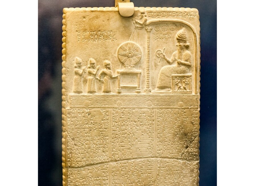
,Title photo description: 29. 07. 2015, LONDON, UK, BRITISH MUSEUM – The Sun God Tablet – 860-850 Bc, Shamash Temple, Sippar, northern Iraq. Ileana_bt – Shutterstock.
SECTION I: INTRODUCTION – „The Electric Temple”
The British Museum, London. Room 55.
Before you hangs a stone tablet not much larger than a sheet of paper. Twenty-nine centimeters high, not quite eighteen wide. Limestone carved in northern Iraq, in Sippar—the ancient city. Two thousand eight hundred and seventy years ago, someone sculpted a scene into this piece of stone that has survived the fall of empires, the forgetting of languages, the shift of religions.
Shamash, the Mesopotamian sun god, sits on his throne. Rays flow from his shoulders. Before him, three figures stand in a posture of adoration, and between them and the deity—a small table with a round solar disk, as if suspended in the air. Symbols, inscriptions, signs whose meaning has been lost in time.
Everyone sees a religious ceremony. An act of worship. Priests before a deity. An ancient ritual captured in stone for eternity.
But there is more.
Look again—this time not at a religious scene, but as a technical diagram. The rectangular shape of the tablet, elongated and layered. The central figure almost mechanical in its symmetry. The rays not decorative, but precisely positioned like wires in a circuit. That hovering table with a circle—not miraculously floating, but supported by clear structural lines. The upper space, the middle section, the base—a layered structure like a cross-section of something real.
Now, imagine you have access to an electron microscope. That you can peer inside every cell of your body. That you see hundreds, thousands of small, elongated structures, layered, with precisely arranged protein complexes. You see how energy flows through them, how protons are pumped, how a molecular turbine spins, producing the fuel of life.
This is a mitochondrion.
And this fragment of stone, no larger than a book page, carved in 860 BC, does not describe a religious ceremony. It describes the most important process in your body—a process our ancestors called the „light of life,” and which we call oxidative phosphorylation. The powerhouse of every living cell.
Welcome to the discovery that changed everything.
SECTION II: FULL SCHEME – „The Complete Map”
April 2022. I’m watching an educational video from Harvard University—”Electron Transport Chain” from the BioVisions series. The animation shows how energy flows through a mitochondrion, how five protein complexes work together to produce ATP. A precise molecular visualization based on years of structural research.
Then I open a photo of the Shamash tablet from the ancient Iraq.
And I see the exact same scene.
„Not loosely. Not if you tilt your head. Not if you really want to believe it. The exact same scene.”
I created a side-by-side—a frame from the Harvard video placed directly below the tablet. I overlaid the same labels on both images. And what was revealed was not just the structure, but the entire flow of energy carved in stone.
, Photo description: 29. 07. 2015, LONDON, UK, BRITISH MUSEUM – The Sun God Tablet – 860-850 Bc, Shamash Temple, Sippar, northern Iraq. Ileana_bt – Shutterstock.
, Video description: Electron Transport Chain, Harvard Online. BioVisions – HarvardX.
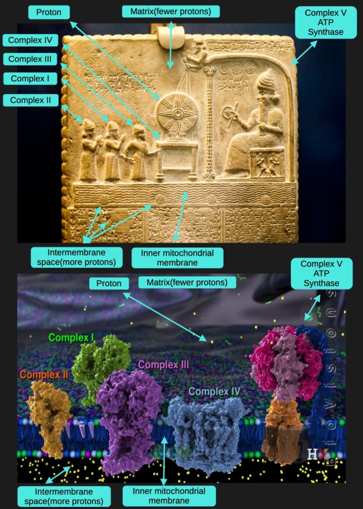
STRUCTURAL MAPPING
The Matrix – The Upper Part (Where It All Begins)
On the Shamash tablet, you see the upper part of the scene—the space above everything, above the complexes, above ATP Synthase.
In the Harvard film, this is the Matrix—the interior of the mitochondrion, the place where the Krebs cycle (also known as the citric acid cycle or TCA cycle – tricarboxylic acid cycle) occurs.
This is where it all begins.
This is where NADH (Nicotinamide Adenine Dinucleotide – reduced form) and FADH₂ (Flavin Adenine Dinucleotide – reduced form) are produced to supply electrons to the entire transport chain.
These molecules are the charged „batteries” full of energy extracted from glucose. NADH will deliver electrons to Complex I, while FADH₂ will deliver electrons to Complex II. Without the Matrix and the Krebs cycle producing these electron carriers, the entire Electron Transport Chain would have nothing to transport.
It is the fuel source for the whole machine.
The Matrix is also where protons will eventually return after flowing through ATP Synthase, completing the cycle.
The Three Figures on the Left – Complexes II, I, III (The Electron Acceptors)
On the Shamash tablet, on the left side, three figures stand in a posture of adoration. Not a random number—exactly three.
The first figure (leftmost) has its hands raised high—a dynamic, announcing gesture, as if proclaiming: „I also contribute!”
The second and third figures (middle and rightmost) stand closer together—their outlines overlap, they are connected.
In the Harvard film, you see three large protein complexes protruding from the mitochondrial membrane:
- Complex II (orange) – first from left
- Complex I (green) – middle
- Complex III (purple) – rightmost
And here’s the crucial detail: Complex I and Complex III are physically connected—they touch, forming a functional supercomplex. Why? Because Complex I transfers electrons directly to Complex III via ubiquinone (Coenzyme Q10), forming an integrated energy transfer system.
And Complex II? The first figure with raised hands is showing: „I also supply electrons to the system!”
Here’s how the electron flow works:
Complex II receives electrons from FADH₂ (produced in the Krebs cycle from succinate oxidation) and delivers them to Coenzyme Q (ubiquinone)—a mobile electron carrier in the membrane.
Complex I receives electrons from NADH (also produced in the Krebs cycle) and also delivers them to the same Coenzyme Q.
Coenzyme Q is like a collection point—gathering electrons from BOTH Complex II and Complex I, then delivering them together to Complex III.
So the electron path is:
- Complex II ← electrons from FADH₂ → Coenzyme Q
- Complex I ← electrons from NADH → Coenzyme Q
- Coenzyme Q → Complex III → (continues downstream)
That’s why the first figure’s hands are raised in that announcing gesture—it’s proclaiming: „I feed electrons into the chain too!” Complex II provides an additional entry point for electrons derived directly from the Krebs cycle.
The critical difference: Complex I and Complex III pump protons across the membrane as electrons flow through them. Complex II does NOT pump protons—it only supplies electrons. But both pathways feed the same downstream electron transport chain.
The tablet shows this perfectly:
- First figure (Complex II) with raised hands = additional electron supplier
- Second and third figures (Complex I and III) touching = physical supercomplex connection
- All three working together, with Coenzyme Q as the mobile shuttle collecting electrons from both entry points
The Electron Handoff – Cytochrome c (The Mobile Messenger)
On the Shamash tablet, look closer at the third figure (Complex III). Its hand extends forward and TOUCHES the table (Complex IV). This is not artistic decoration—this is molecular precision.
In biochemistry, Complex III transfers electrons to Complex IV through cytochrome c—a small, mobile protein that physically shuttles between them through the intermembrane space.
In the Harvard film, cytochrome c is shown as a molecular messenger carrying electrons from Complex III to Complex IV. It physically moves, detaches from Complex III, travels through the intermembrane space, docks at Complex IV, and delivers its electron cargo.
The hand of Complex III touching the table represents this electron carrier in action. The physical touch on the tablet = the molecular handoff happening continuously.
Cytochrome c literally „hands off” electrons from Complex III to Complex IV, exactly as the carved gesture shows.
Another detail of perfect functional accuracy. The ancients understood not just the static complexes, but the dynamic transfer between them—the mobile carriers that make the system work.
The Table with the Circular Disk – Complex IV + Regulatory Control (The Final Proton Pump)
On the Shamash tablet, between the three figures and the deity, a table with a circular solar disk appears to hover. Suspended, with structural ropes running upward, connecting the table to two small figures at the end of a structural element extending from behind the central deity.
In the Harvard film, you see Complex IV (blue/grey)—the final complex in the electron transport chain. This is where electrons complete their journey, being transferred to oxygen to form water (H₂O). But more importantly for ATP production, this is where the final protons are pumped across the membrane.
The circular disk on the table is a MAGNIFIED PROTON—a pedagogical detail showing exactly what is being pumped.
Notice carefully: The circular disk has the IDENTICAL pattern as the small spheres (protons) flowing through the waves below—only magnified, enlarged to show detail. The ancients are demonstrating: „These small spheres throughout the structure? They are protons. Here’s what one looks like up close, in detail.”
This is brilliant pedagogy—showing both the many small protons in motion AND a magnified example so you understand what you’re looking at.
And the ropes connecting upward? They show REGULATORY CONTROL—the feedback mechanism.
The two small figures at the end of the peripheral stalk (extending from ATP Synthase) hold ropes leading down to Complex IV. This represents respiratory control—the sophisticated feedback system where ATP Synthase regulates the entire electron transport chain.
When ATP Synthase slows down (due to insufficient ADP or other factors), the proton gradient builds up to maximum. When the gradient is too high, Complex IV cannot pump more protons against it—the system must slow or stop. The peripheral stalk literally „holds Complex IV in check” through this regulatory feedback.
The tablet shows functional feedback carved in stone: ATP Synthase → controls → Complex IV.
This is molecular engineering—a control circuit ensuring the system runs at the right speed, neither too fast nor too slow.
The Inner Mitochondrial Membrane – The Lower Waves (The Barrier)
On the Shamash tablet, you see undulating patterns resembling ocean waves. Within and around these waves, you see small, round spheres flowing, moving through the undulating structure.
In the Harvard film, you see the inner mitochondrial membrane, which is folded into structures called cristae. These waves on the tablet are an exact representation of the folded membrane—not decorative, but structurally accurate.
And those small spheres? They are protons—shown in the film as yellow particles.
This is the barrier. The inner membrane separates two spaces and is relatively impermeable to protons. Complexes I, III, and IV pump protons across this membrane, from the Matrix (above) into the Intermembrane Space (below).
The membrane is the foundation of the proton gradient—without this barrier, protons would diffuse freely and no gradient could form.
The waves are not decoration—they are precise structure. The spheres are not symbols—they are protons in motion, being pumped, accumulating, creating the pressure that will drive ATP Synthase.
The Intermembrane Space – Beneath the Waves (The Pressure Chamber)
On the Shamash tablet, below the undulating patterns (the inner membrane), you see another layer—a distinct space, an area beneath the waves. This is where those small spheres (protons) accumulate after being pumped by the complexes.
In the Harvard film, you see the intermembrane space—the area between the inner and outer mitochondrial membranes.
This is the proton reservoir.
As Complexes I, III, and IV pump protons across the inner membrane, those protons accumulate here, creating a concentration gradient. The proton concentration is HIGH in the intermembrane space and LOW in the Matrix.
This creates electrochemical potential—stored energy, like water behind a dam. The protons „want” to flow back into the Matrix, but the inner membrane blocks them. They can only return through one route: ATP Synthase.
This is where the proton „pressure” builds up that will drive the molecular turbine. It is an energy reservoir—protons wait, accumulate, build potential, creating the force that will spin the rotor of ATP Synthase and produce ATP.
The greater the difference in proton concentration between the intermembrane space and the Matrix, the stronger the driving force for ATP production.
The Central Figure (Shamash) – Complex V, ATP Synthase (The Energy Transformer)
On the Shamash tablet, centrally and on the right, the deity sits on a throne. The largest figure. The most ornate. The most detailed. Majestic, controlling—the culmination of everything leading up to this point.
In the Harvard film, on the right side, a massive structure dominates—shown in pink-purple, orange, and navy blue. This is ATP Synthase—Complex V—the molecular turbine that transforms the proton gradient into ATP.
This is the final step.
All the electron transport, all the proton pumping, all the gradient building—it all exists for this moment. The accumulated protons in the intermembrane space flow back through ATP Synthase into the Matrix, and as they flow, they drive the rotation of a molecular turbine spinning at 9,000 RPM.
This rotation produces ATP—the universal energy currency of life.
ATP Synthase is where energy transformation happens. The electrochemical potential of the proton gradient becomes the chemical bond energy of ATP.
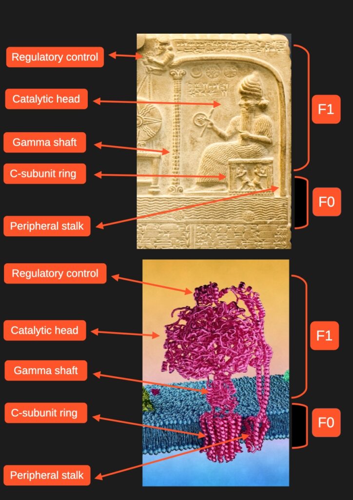
The detailed structure of this remarkable molecular machine—its catalytic head, gamma shaft, c-ring rotor, peripheral stalk, and regulatory components—will be described in detail in Section III: „Shamash – The Molecular Sun God.”
But Wait—Where Are the Electrons?
The Harvard film is titled „Electron Transport Chain”—it shows blue/cyan dots (electrons) hopping between complexes: Complex I → Coenzyme Q → Complex III → cytochrome c → Complex IV → oxygen.
But the Shamash tablet shows primarily protons—the small yellow spheres in the waves, the magnified disk on the table.
Why this difference?
Because the ancients understood something profound: electrons are the MEANS, protons are the RESULT.
Here’s what happens in the Electron Transport Chain:
Step 1: Electrons (blue in the film) flow through Complexes I, II, III, and IV
Step 2: This electron flow POWERS the pumping of protons (yellow) across the membrane
Step 3: Protons accumulate in the intermembrane space, creating a gradient
Step 4: This proton gradient DRIVES ATP Synthase
Step 5: ATP is producedThe electrons (blue) are the transfer mechanism—invisible energy carriers that hop between protein complexes, transferring energy extracted from NADH and FADH₂. They enable the pumping, but they themselves are not accumulated or stored.
The protons (yellow) are the currency of stored energy—the visible, measurable result. They accumulate, creating pressure. They flow through ATP Synthase. They are what directly drives the rotor. They are the intermediate form of energy between electron transport and ATP synthesis.
The ancients chose to show what drives the final machine—the protons—rather than the electron transfer mechanism between complexes.
This is a pedagogical choice, a decision about what’s most important to show. Just as modern biochemistry textbooks often focus on the proton-motive force and proton gradient when explaining ATP synthesis, rather than dwelling on electron orbital mechanics.
The tablet shows the FUNCTIONAL CURRENCY (protons), not just the transfer mechanism (electrons).
It’s like showing water flowing through a turbine (protons driving ATP Synthase) rather than showing the pump that moved the water there (electron transport). Both are necessary, but one is more directly relevant to understanding the final energy transformation.
Notice also: The magnified disk on Complex IV’s table could represent both the proton being pumped AND the endpoint where electrons meet oxygen. A dual symbol showing where the electron journey ends (e⁻ + O₂ → H₂O) and where the final protons are pumped.
But the emphasis is clearly on protons—because protons are what Shamash (ATP Synthase) consumes to produce ATP.
In the film, you can see electrons (blue) entering Complex I and Complex II, traveling through Coenzyme Q to Complex III, then through cytochrome c to Complex IV, and finally to oxygen. But simultaneously, you see protons (yellow) being pumped, accumulating, and eventually flowing through ATP Synthase.
The ancients understood that the electron transport chain exists TO CREATE the proton gradient. They showed the end result that matters most: the accumulated protons (yellow) that power life’s most important machine.
THE COMPLETE PICTURE
Everything fits. Element by element. Structure by structure. Placement by placement. Function by function.
From top to bottom, from beginning to end:
Matrix → produces NADH and FADH₂
↓
Complexes II, I, III → accept electrons, pump protons
↓
Cytochrome c → shuttles electrons from III to IV
↓
Complex IV → completes electron transport, pumps final protons, regulated by ATP Synthase
↓
Inner Membrane → the barrier creating separation
↓
Intermembrane Space → proton reservoir, high concentration
↓
ATP Synthase → protons flow through, producing ATP, returning to MatrixThis is not a coincidence. This is not „seeing patterns everywhere.” This is a precise representation of a molecular process in a form the ancients could carve in stone—a 2D functional schematic showing internal structures and energy flow.
This is not a religious ceremony carved in stone.
This is a ceremony of ENERGY carved in stone.
The flow of electrons from NADH and FADH₂, carried through Coenzyme Q and cytochrome c. The pumping of protons creating a gradient. The regulatory feedback from ATP Synthase to Complex IV. The spinning rotor driven by returning protons. The production of ATP—all carved with functional precision.
Every element of the tablet describes a step in this molecular ceremony. A step in transforming the energy of the sun (captured by plants, eaten by you, broken down in the Krebs cycle) into ATP—the energy your cells can actually use.
Watch the Harvard film. See how the electrons (blue) flow from complex to complex through mobile carriers. See how the protons (yellow) are pumped across the membrane, accumulating in the intermembrane space. See how they flow back through ATP Synthase, spinning the rotor. See the entire ceremony in action—the exact same ceremony carved on the tablet from 860 BC.
Then look at that tablet.
And tell me it’s a coincidence.
SECTION III: SHAMASH – THE MOLECULAR SUN GOD
In the temple of Ebabbara—the priests carved life’s greatest secret into limestone. They named it Shamash, the Mesopotamian sun god. But Shamash was not just a god of the sun in the sky. Shamash was the god of ENERGY—the energy that flows from the sun through plants, through food, all the way to the molecular machine in every cell.
And this machine is exactly what they carved in stone.
,Photo description: 29. 07. 2015, LONDON, UK, BRITISH MUSEUM – The Sun God Tablet – 860-850 Bc, Shamash Temple, Sippar, northern Iraq. Ileana_bt – Shutterstock
, Illustration description: 3d illustration of the ATP synthase. Meletios Verras – Shutterstock.
THE COMPLETE STRUCTURE OF ATP SYNTHASE
Let’s examine the central figure on the tablet—Shamash sitting on the throne. Every detail of this carved deity corresponds to a specific component of the most complex molecular machine known to science.
F1 – THE CATALYTIC HEAD (Upper Part)
On the tablet: The seated figure of Shamash—the upper body, head, torso. Majestic, immobile, controlling.
In biochemistry: The F1 catalytic head consists of the α₃β₃ hexamer—three alpha and three beta subunits arranged in a ring. This is where the miracle happens: where ADP and phosphate are joined to form ATP.
The F1 head remains structurally stable while the gamma shaft rotates inside it. Each rotation causes conformational changes in the beta subunits, cycling through three states:
- Open (empty, ready to receive substrates)
- Loose (ADP + Pi loosely bound)
- Tight (ATP tightly bound and released)
This is the „factory” where ATP is manufactured—approximately 100-150 molecules per second per machine.
And what Shamash holds in his hand? A circle and a rod—the classic Mesopotamian „ring and rod” symbols of power.
But look closer. This is not just ATP as a finished product. This is a hydrogen bond.
The circle and rod represent the molecular mechanism of joining—the hydrogen bonds that hold ADP and phosphate in position before the chemical reaction occurs. Hydrogen bonds are the invisible bridges that enable catalysis. Without them, no ATP would be produced.
The ancients understood not just the product, but the process—the molecular mechanism at the level of chemical bonds.
GAMMA SHAFT (Central Column)
On the tablet: A visible central column running vertically through the middle of Shamash, from the throne base upward.
In biochemistry: The gamma (γ) shaft is the central rotating axle of ATP Synthase. It extends from the c-subunit ring (F0 rotor) through the center of the F1 catalytic head.
As protons flow through F0, they cause the gamma shaft to spin at 9,000 revolutions per minute—faster than a Formula 1 engine. This rotation drives the conformational changes in the F1 catalytic head that produce ATP.
Why is it visible on the tablet? Because the tablet is a 2D functional schematic—a cross-section, like an engineering blueprint. In 3D visualizations from outside, the gamma shaft is hidden inside the catalytic head. But on the tablet, it’s shown as a visible element because this is a pedagogical diagram revealing the internal architecture.
The ancients created a cutaway view—exactly as we do in modern biochemistry textbooks when we need to show how the machine works internally.
C-SUBUNIT RING (The Throne + Rotating Base)
On the tablet: The throne on which Shamash sits, and the base with two small figures in dynamic, spinning poses.
In biochemistry: The c-subunit ring is the F0 rotor—a ring of 8-15 c-subunits (depending on species) embedded in the inner mitochondrial membrane. This is what actually rotates.
Protons flow through channels between the c-ring and the a-subunit, causing the c-ring to spin. The c-ring is connected to the gamma shaft, so when it rotates, the gamma shaft rotates—driving ATP production in the F1 head above.
The two dynamic figures at the base? They perfectly capture the spinning motion—caught mid-rotation, like dancers frozen in a whirl. This is the fastest-spinning component in biology.
Shamash sits ON this rotating throne—showing that the F1 head (Shamash) is mechanically coupled to the rotating base (c-ring rotor).
PERIPHERAL STALK (Behind Shamash)
On the tablet: Structural elements visible behind Shamash’s back, extending from the base upward. At the top of this structure, two small figures hold ropes leading to Complex IV.
In biochemistry: The peripheral stalk (also called the b₂ complex) is a long, stable protein structure that runs alongside the gamma shaft from F0 to F1. Its crucial function is to prevent the entire F1 head from rotating—it acts as a stator, a stabilizer.
Without the peripheral stalk, when the gamma shaft spins, the entire F1 head would spin with it—no ATP would be produced. The peripheral stalk holds F1 stable while allowing only the gamma shaft to rotate inside.
The two figures at its end holding ropes? This shows the regulatory control mechanism. The peripheral stalk doesn’t just stabilize—it also extends to regulate Complex IV through respiratory control. When ATP Synthase slows down, proton gradient builds up, and Complex IV must slow or stop. The tablet shows this functional feedback connection. THE THREE-ION CODE (wersja zwięzła, bez kodu)
THE THREE-ION CODE
In the upper section of the tablet, beside the central column, there is a rectangular panel containing three symbols: one crescent and two perfect disks.
Archaeologists interpret them as celestial symbols: Moon, stars, sun.
But they represent something far more precise.
The rectangular panel is the intermembrane space—the compartment where protons accumulate. The three symbols represent the ionic components of ATP synthesis:
• The crescent ☽ = Magnesium ion (Mg²⁺)
Charge: +2. Catalytic cofactor. ATP cannot exist without it.• Two disks ⚫⚫ = Protons (H⁺)
Charge: +1 each. Energy source driving rotation.The crescent has a different shape because magnesium has a different charge and function than protons.
They knew the gradient. They knew the ions. They knew the chemistry.
2,870 years before Mitchell’s Nobel Prize (1978). Before we discovered ATP-Mg²⁺ complexes. Before we solved the crystal structure.
REGULATORY CONTROL (Top)
„The two figures holding ropes show the regulatory control mechanism connecting to Complex IV (described in Section II).”
WHAT WE STILL DON’T KNOW
Here’s something remarkable: Despite Nobel Prizes awarded in 1997 and 2019 for ATP Synthase research, we still don’t fully understand every detail of this machine.
In the Harvard BioVisions films, the narrators explicitly state: „Many structural details remain unknown” and „Some components are still being discovered.”
What we know:
- The basic structure of F1 and F0
- The rotational mechanism
- The approximate positions of major subunits
What remains partially mysterious:
- Exact dynamics of rotation in real-time
- Precise coupling mechanism between proton flow and rotation
- Full 3D structure of all conformational states
- Functions of some regulatory subunits (IF1, subunits e, f, g)
- Complete understanding of how different organisms vary their ATP Synthase
The ancients carved this machine in 860 BC. We’re still discovering its details in 2025.
How did they know?
FROM SUN TO LIFE – THE COMPLETE CYCLE
Why a sun god specifically? Because the ancients understood the complete energy cycle:
Sun → photons → Plants → photosynthesis → glucose → Animals → eat plants → Mitochondria → break down glucose → Electron Transport Chain (Complexes I-IV) → transfer energy → ATP Synthase (Shamash) → transforms into ATP → LIFE
All life’s energy originates from the sun. But the transformation of that energy into usable cellular fuel happens inside—in the mitochondria, in ATP Synthase, in Shamash.
They chose the final link—ATP Synthase—as the representation of the entire process. They named it Shamash, the sun god, because it is precisely solar energy—after passing through the entire chain—that concludes its journey in this spinning turbine.
They KNEW.
A MACHINE, NOT A METAPHOR
Shamash is ATP Synthase—Complex V. When I say „machine,” I am not using metaphor. It is literally a mechanical turbine with spinning parts, axles, a stator, a rotor—only built from proteins instead of metal.
Imagine a water turbine: Water flows from top to bottom → sets a wheel in motion → wheel turns an axle → axle drives a generator → generator produces electricity.
ATP Synthase works exactly the same way:
Protons flow from intermembrane space into matrix → flow through F0 rotor → rotor spins at 9,000 RPM → gamma shaft rotates inside F1 → conformational changes produce ATP.
It is a molecular power plant.
AN UNFATHOMABLE SCALE
ATP Synthase produces about 100-150 ATP molecules per second. In a single one of your cells, there may be 2,500 mitochondria. Your body contains trillions of cells.
Do the math.
Your body produces and uses its own body weight in ATP every day—about 70 kilograms of ATP daily. This is not an exaggeration. It is a reality measured, calculated, documented.
ATP is the energy currency of life. Every muscle movement, every thought in your brain, every DNA repair, every protein synthesis—all require ATP. And ATP Synthase is the only bank that produces it.
THE SHAMASH THAT NEVER SETS
The ancients said that Shamash „travels through the underworld at night” to rise again in the morning. Most historians think this is poetry, a metaphor.
It is not poetry. It is molecular truth.
ATP Synthase works 24/7. While you sleep and while you are awake. Day and night. It „travels through the underworld” (the mitochondrial matrix), transforming energy even after the sun has set, even when your eyes are closed, even while you dream.
Shamash was described as „the one who sees all.” The true Shamash—ATP Synthase—truly sees all. It is in every cell. In every tissue. In every organ.
WHY THEY CHOSE THIS MACHINE
Why did the ancients carve ATP Synthase? Why preserve THIS specific molecular process in stone?
Because it is the most universal, most fundamental process of life on Earth.
Not every organism has eyes. Not every one has a heart. Not every one has a brain. But every living organism that requires oxygen has ATP Synthase.
- Plants have ATP Synthase (in mitochondria and chloroplasts)
- Animals have ATP Synthase
- Fungi have ATP Synthase
- Protists have ATP Synthase
From amoeba to elephant, from algae to sequoia—everyone has Shamash.
This is the foundation of life. Without energy—there is no protein synthesis, no DNA replication, no cell division, no life.
Perhaps this is why ATP Synthase was chosen to be preserved in stone. As a fundamental truth worth surviving millennia:
Life is energy. And energy is transformed by a molecular machine that never stops working.
Whether this was the first biological process the ancients encoded, or one of many, we may never know. But the choice to preserve THIS process—the most universal energy transformer in all of life—reveals their understanding of what matters most.
SIPPAR – THE CITY OF KNOWLEDGE
Sippar was not a random location. It was a city built as a cell—with the temple of Ebabbara representing the nucleus, the command center.
The temple’s name: Ebabbara—”The Luminous House,” „House of Light.”
Not „house of prayer.” Not „house of sacrifice.” House of Light.
And inside this temple-nucleus, a small limestone tablet (29cm × 18cm) preserved the schematic of mitochondria—the tiny powerhouses of the cell.
The hierarchy mirrors cellular structure:
- Sippar = The complete cell
- Ebabbara temple = The nucleus (~10-20 μm, largest organelle)
- Shamash tablet = Documentation of mitochondria (~1 μm, much smaller)
Why is mitochondrial documentation kept in the nucleus-temple?
Because this is exactly how cells work. The nucleus contains DNA with instructions for building mitochondria. Nuclear DNA encodes most mitochondrial proteins. The nucleus is the archive preserving blueprints for all cellular components—including the energy-producing mitochondria.
The small tablet inside the massive temple reflects the true scale: mitochondria are tiny organelles compared to the nucleus. Yet this small tablet describes the most fundamental process—energy transformation happening in thousands of powerhouses distributed throughout the cellular city.
Ebabbara – House of Light – the nucleus preserving knowledge of how cellular powerhouses work, exactly as nuclear DNA preserves the code for mitochondrial machinery.
THEY KNEW
The precision carved into this limestone was no accident. The ancient Mesopotamians deliberately encoded this knowledge.
They knew that the true Shamash, the true source of life’s light, was not only in the sky but inside the cells. They knew that solar energy must be transformed by a molecular machine. They knew what this machine looked like—the catalytic head, the gamma shaft, the rotating base, the peripheral stalk, the regulatory control.
And they carved this truth in limestone—the complete schematic of ATP Synthase—so that we could discover it 2,870 years later when we would be ready to understand it.
, Video YouTube ATP synthase in action – Harvard Online. Molecular Machines – Atp Synthase it’s been called one of the wonders of the molecular World an amazing nanoscale machine.
SECTION IV: SIDE VIEW – „The Stone That Breathes”
Step back from the tablet. Stop reading the symbols, stop analyzing the figures. Just look at the shape of the stone itself from the side.
You see an elongated block of limestone. But it is not a simple, evenly carved rectangular prism. Look at its thickness—its side profile. It is not uniform across its entire length.
At the top—a narrowing.
At the center—a widening, maximum thickness.
At the bottom—another narrowing.An organic shape. As if the stone were „breathing,” expanding in the middle and tapering at the ends. A rounded contour not only from top to bottom but also in depth—swelling at the center, narrowing at the edges.
But there is more. Look carefully at the side of the tablet.
, Photo description: Limestone tablet. LONDON, UK, BRITISH MUSEUM – The Sun God Tablet – 860-850 Bc, Shamash Temple, Sippar, northern Iraq. The British Museum.
, Photo description: Mitochondria are visible under the light microscope although little detail can be seen. Barou abdennaser – Shutterstock.
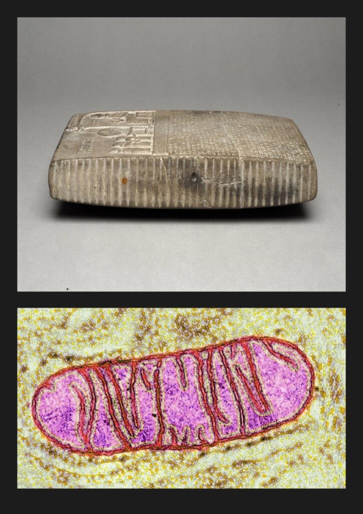
You see regular grooves. Vertical striations running across the width of the stone. These are not random marks of time—they are deliberately carved lines, equally spaced, regular, running across the entire side profile. Like transverse incisions, like ridges.
Now look at a photo of a mitochondrion under a microscope.
Exactly the same pattern.
A mitochondrion is not smooth inside. It has cristae—internal folds of the mitochondrial membrane that look like transverse ridges, like combs running across the length of the organelle. These cristae are particularly visible under an electron microscope—regular, equally spaced structures running across the width of the mitochondrion.
And exactly the same pattern is visible on the side of the Shamash tablet. Regular grooves running transversely. Cristae carved in limestone.
This is not a flat slab with a relief. This is a three-dimensional model of a mitochondrion carved in stone—with an organic external shape (widening in the center, tapering at the ends) and with an internal cristae structure (transverse grooves).
In 1857, Rudolf Virchow, looking through a light microscope, saw these organic shapes in cells. Wider in the center, narrower at the ends. He called them „granules.” In 1898, Carl Benda named them „mitochondria”—thread-like grains.
But the cristae? Those transverse structures inside? They were not seen until the electron microscope in the 1950s. George Palade received the Nobel Prize in 1974 for describing the ultrastructure of mitochondria, including the cristae.
And on the Shamash tablet, the cristae have been carved since 860 BC.
„Regular grooves running across the tablet’s side. Cristae carved in limestone.
This changes everything.
Because if they could see cristae—the internal folds of mitochondrial membranes—then they had tools to observe cellular ultrastructure. And if they had such tools, they could see everything carved on the front of the tablet:
- ATP Synthase spinning at 9,000 RPM
- Protons flowing through membrane channels
- Electron Transport Chain complexes pumping energy
- The molecular machinery of life
The cristae aren’t the mystery. They’re the confirmation.
Confirmation that someone possessed the technology to observe mitochondria in detail. To understand their internal architecture. To document their molecular machines.
In 1950, George Palade saw cristae for the first time through an electron microscope. He received the Nobel Prize in 1974.
The cristae were carved in stone 2,810 years earlier.
Someone knew. Someone saw. Someone documented.”
, Video You Tube – Mitochondria: the cell’s powerhouse – Harward Online
SECTION V: INSIDE THE POWERHOUSE – „Cristae Cathedral”
We’ve seen the tablet. We’ve seen the schematic. We’ve seen how it works.
Now let’s go inside.
Imagine you are inside a mitochondrion. You’re not looking at it from the outside through a microscope—you are THERE. Inside. In the Matrix, surrounded by cristae—those folded inner membranes that look like the majestic columns of a cathedral.
,Video YouTube – Mitochondria interior – Transection through cristae tubule (2020) Drew Berry wehi.tv
Drew Berry’s film, „Mitochondria Interior – Transection through cristae tubule,” shows exactly that. The camera show animation the interior of a mitochondrion. You see gigantic folds of the cristae membranes jutting out like canyon walls. You see two on left and two on right of ATP Synthases—those pink-purple turbines—protruding from every cristae like mushrooms, like a forest of molecular machines.
You are standing inside a molecular power plant. Not a metaphorical one—a literal one. Around you, the turbines spin at racing engine speed, pumping out energy, producing the fuel of life.
And this is just ONE mitochondrion.
A single cell may contain 2,500 such mitochondria.
Your body contains trillions of cells.
Stop. Try to grasp this with your mind.
Every second of your life—whether you’re sleeping, running, or reading these words—trillions of mitochondria, each containing hundreds of ATP Synthases, all spin simultaneously. Billions of molecular turbines working in synchrony, producing your body weight in ATP every day.
70 kilograms of ATP. Daily. This is not an exaggeration—it is a reality measured, calculated, documented.
Now look again at the Shamash tablet.
This piece of limestone, no larger than a book page, describes a process that is happening right now in every cell of your body. In the muscle cells of your heart, beating right now. In the neurons of your brain processing these words. In the cells of your liver, kidneys, skin, eyes.
Everywhere. All the time. From the moment of conception to your final breath.
This ceremony carved on the tablet is not an ancient ritual that ended.
This ceremony is happening NOW. In you. In me. In every living organism on this planet.
Mitochondria are in the cells of plants, animals, fungi. Bacteria have similar systems. The same fundamental machine—electron transport chain + ATP Synthase—operates in every life form that requires energy.
Life on Earth is powered by this molecular turbine. By this spinning machine that the ancients called Shamash—the god of the sun, the god of light, the god of energy.
Because they were right.
„The energy of the sun—through the complete cycle described in Section III—culminates in this spinning rotor.”
Shamash is not a myth.
Shamash is the actual molecular machine in every one of your cells. Spinning 9,000 times a minute. Producing the energy of your life.
And when I watch Drew Berry’s film, when I see those of ATP Synthases protruding from the cristae like a forest of molecular machines, when I try to grasp with my mind that this is all happening in billions of places simultaneously within my own body—I realize how advanced life on Earth is.
When I look at these films and images, I see proof of intelligent design.
And I love You, my God!
Thank You for this design. For this machine. For this life.
Thank you to the creators of these films—Robert Lue, Dale Muzzey from Harvard BioVisions. Drew Berry from WEHI. Without your work, without these visualizations, this discovery likely would never have been made. At least not by me. You have shown the world the work of God the Creator.
Thank you to the photographers from Shutterstock—Ileana_bt, Meletios Verras, Barou Abdennaser. Through your lenses, you captured the design of God the Creator.
Thank you to the British Museum in London for preserving this tablet.
Thank you all!
SECTION VI: „THE DISCOVERY – When Two Worlds Met”
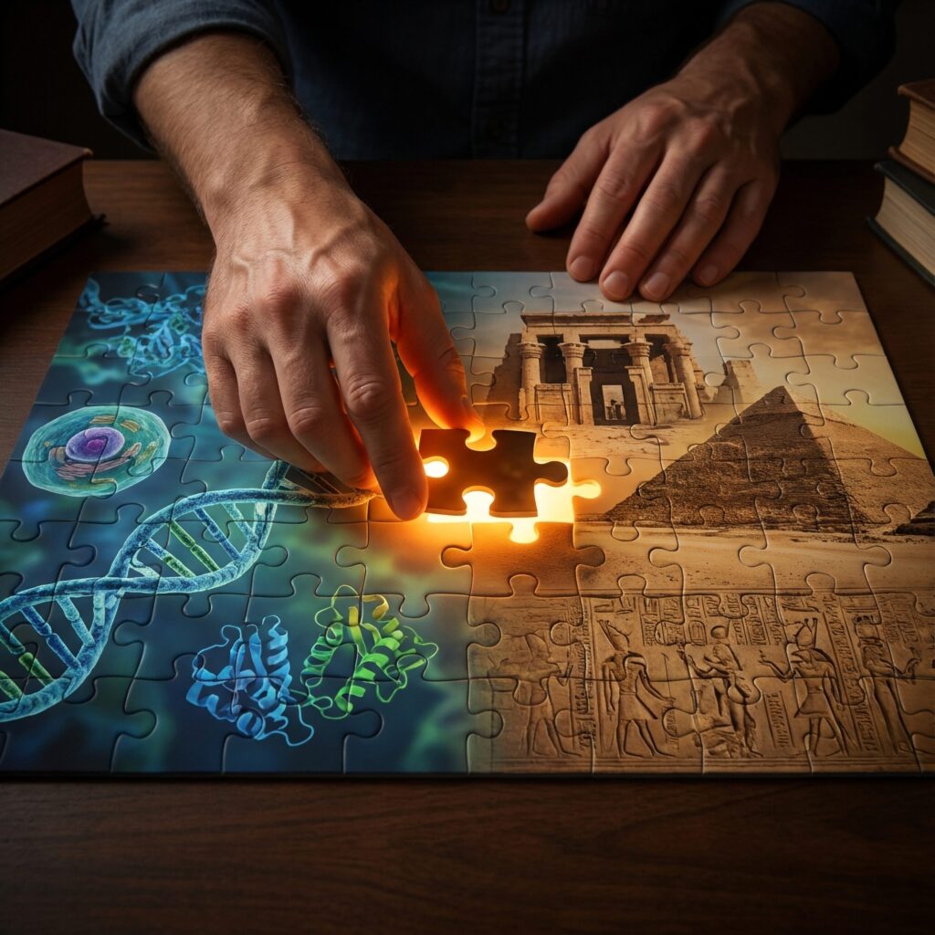
,image description:Hands placing last puzzle piece with light glow, puzzle shows molecular biology on left (DNA, cells, proteins) and ancient architecture on right (temples, carvings, monuments), high quality.Shutterstock AI.
Year 2022. I have two parallel interests consuming my free time.
On one hand—molecular biology. I watch films about cells, proteins, life processes at a level invisible to the naked eye. I am fascinated by the precision, complexity, and intelligence of life’s design at the molecular level.
On the other hand—the architecture of the old world. Lost Tartaria. Old photographs of cities from years ago. Buildings that no longer exist, destroyed in wars, burned, demolished. Cathedrals, palaces, temples, monuments, art, painting, sacral symbolic—all in the same style, scattered across the world. Europe, Asia, both Americas. Everywhere, the same architectural language.
People try to explain it in various ways. Some say it’s an alien civilization. Others claim that cathedrals were power plants using atmospheric energy. Still others search for free energy, hidden technologies, erased traces of an advanced civilization.
I look at the same photographs. And I begin to see something else.
THE MOMENT OF CONNECTION
One day, I’m watching a film: „Electron Transport Chain – Harvard Online, BioVisions – HarvardX”.
It’s a beautiful animation of mitochondrial energy production. Complexes I, II, III, IV. ATP Synthase spinning. Proton gradient. The precision of a molecular machine.
Suddenly, looking at this arrangement—four complexes, the central ATP Synthase—my brain makes a strange connection.
This looks like an architectural schematic.
Not „vaguely resembles.” It has the structure of a design. A composition. An order I see in old architectural drawings.
And then I remember something. I’ve seen a similar layout somewhere. Not in building architecture, but in…
Mesopotamian tablets.
THE SHAMASH TABLET
I google: „Mesopotamia sun god tablet Shamash”.
And I find: Tablet of Shamash. British Museum. 860 BC. Sippar, Iraq.
I look at a photo of this limestone tablet.
And my world stops.
Three figures on the left—Complexes I, II, III touching and connected. One hand of Complex III extending to touch the table—cytochrome c transfer. Table with a circular disk—Complex IV with a magnified proton. Ropes from the table upward—regulatory control mechanism. The large central figure Shamash—ATP Synthase. Waves at the bottom—mitochondrial membranes with protons.
This is exactly the same layout as in the Harvard film.
Element by element. Position by position. Function by function.
THE SCHEMATIC UNFOLDS
I sit for hours comparing. I take screenshots. I highlight elements. I check details.
And I begin to see more:
The seated Shamash = F1 catalytic head (where ATP is made)
The throne beneath him = C-ring rotor (the spinning base)
The visible central column = Gamma shaft running through the center
Wait. In the 3D film, the gamma shaft is INSIDE the catalytic head—invisible from outside. But on the tablet, it’s shown as a visible column.
Then I understand: The tablet is a 2D cross-section. A functional schematic. Like an engineering blueprint that shows internal components normally hidden from view.
The ancients created a cutaway diagram—exactly as we do in modern textbooks when we need to show how a machine works internally.
Structures behind Shamash’s back = Peripheral stalk (stabilizer)
Two figures at the top holding ropes = Regulatory control extending to Complex IV
Two dynamic figures at the base = The spinning c-ring captured in motion
The circle and rod in Shamash’s hand = Not just ATP, but a hydrogen bond—the molecular mechanism of joining ADP and phosphate
This is not a religious scene. This is a technical drawing of a molecular machine with its complete functional architecture—catalytic head, rotating shaft, spinning base, peripheral stalk stabilizer, regulatory feedback system.
And it’s a 2D schematic that reveals internal structures we can only see in cross-section.
WHAT THIS MEANS
The ancients were not primitive. They were more advanced than us.
They had knowledge of molecular biology. They had tools—I don’t know what kind, but since they could carve an exact schematic of ATP Synthase including internal structures like the gamma shaft, they had a way to see and understand it.
They understood the difference between 3D external appearance and 2D functional schematics. They chose to carve a cross-section—a pedagogical diagram—exactly as we do in modern science education.
And they encoded this knowledge. In architecture. In symbols. In tablets. In monuments.
Not because they lacked books or notebooks. But because stone survives. Stone survives wars. Fires. Floods. The fall of civilizations. Oblivion.
They carved their knowledge in stone so that future generations—us—could find it when we were ready to understand it.
THE FUNDAMENTAL QUESTION
But if they knew—if they had this knowledge of molecular biology, of cell structure, of internal machine components—then how did we forget?
How could a civilization that could carve a 2D cross-section of ATP Synthase showing the gamma shaft, c-ring rotor, and peripheral stalk in 860 BC lose this knowledge so completely that we only began to rediscover it in the 20th century?
What happened? Wars? Catastrophes? Deliberate destruction of knowledge?
I look at old photographs of burned cathedrals, demolished palaces, destroyed libraries. I see the systematic destruction of old architecture over the last 200 years. World War I. World War II. Revolutions. Arson. Demolitions „for progress.”
Was it accidental? Was someone deliberately destroying evidence?
These are questions I will try to answer in future posts. This is a deeper story than just one tablet from Sippar. This is a story of lost knowledge, an advanced civilization, and encoded instructions left in stone.
THE BEGINNING OF THE BLOG
But that day in April 2022, looking at the Shamash tablet and the Harvard film side by side, I knew one thing:
I must document this.
However, two years passed before I founded CellGod.live. I needed time to gather more evidence. Find more connections. Understand the deeper pattern.
Finally—January 6, 2025—I publish the post about the Shamash tablet as a mitochondrion. It was one of the first articles on the blog.
But back then, I couldn’t describe it well yet. I saw the identical layouts, but I didn’t yet have the full language to express it.
So now – almost a year later – I’m returning to this post. I’m refining its message.
Because over these months I’ve written over 58 articles. I’ve discovered dozens of connections:The biblical Ark of the Covenant as RNA Polymerase I. Artemis of Ephesus as the proteasome. The Warsaw Mermaid as protein complexes. Jesus Christ as RNA—mRNA, tRNA, rRNA—the ribonucleic acid that carries divine instructions. Christmas traditions as the encoded schematic of pre-mRNA formation.
And sacred sites around the world as types of cells:
Vietnam → Basic animal cell
Alabama → Plant cell
Russia → Neutrophil (immune cell)
Germany → Monocyte (phagocytic cell)
Netherlands → Human skin cell
Portugal → Fibroblast (connective tissue)
Malaysia → Human cell (organelles)
Kazakhstan → Melanocyte (pigment cell)Everything you see on CellGod.live has its origin in that film—”Electron Transport Chain.”
From the moment my two worlds—molecular biology and ancient architecture—met at a single point: the Shamash tablet.
From the moment I understood that not only was life designed at the molecular level, but someone left us instructions. Encoded. Carved in stone. As functional schematics showing internal architectures. Waiting until we were ready to read them.
And that day in April 2022, I began to read them.
Thank You my God Creator for giving me eyes to see this work. For connecting these two worlds in my mind. For allowing me to recognize the schematic that survived for 2,870 years.
SECTION VII: „IMPOSSIBLE KNOWLEDGE – The Timeline of Impossibility”
Let’s lay out the facts. Let’s examine the chronology of scientific discoveries and ask the question that demands to be asked:
How is this possible?
860 BC – The Tablet Is Carved
Sippar, northern Iraq. During the reign of King Nabu-apla-iddina (888-855 BC).
A craftsman carves into a limestone tablet (29.21cm × 17.78cm):
- Three electron transport complexes (I, II, III)
- Complex IV with an electron above it
- ATP Synthase with stator, rotor, peripheral stalk
- Proton gradient (spheres in waves)
- Mitochondrial membranes (waves)
- Intermembrane space (space below the waves)
- ATP product (circle + rod in hand)
Everything is precise. Everything is functional. Everything exactly where it should be.
2,870 years ago.
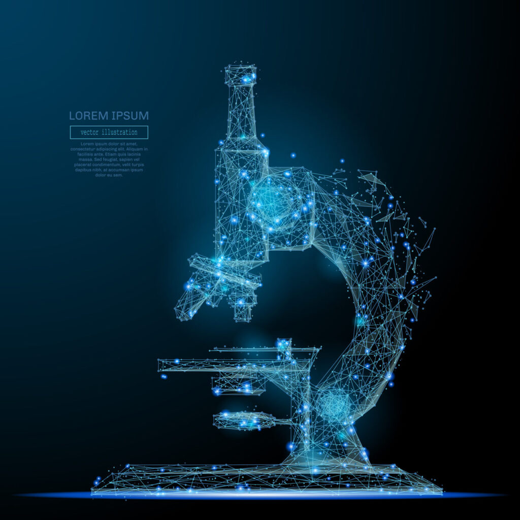
,Vector description: An abstract microscope image in the form of a starry sky or space, consisting of points, lines and shapes in the form of planets and the universe. Vector education and skeletal concept of science. AntonKhrupinArt – Shutterstock.
Someone SAW this. Someone UNDERSTOOD it. Someone had the tools and knowledge to observe molecular structures and carve them in stone.
Now let’s see when we „officially” discovered what is carved on this tablet.
1857 – The First „Granules”
Rudolf Virchow looks through a light microscope at cells. He sees small bodies inside. He calls them „granules.”
He doesn’t know what they are. He doesn’t know their function. He only sees shapes.
2,717 years after the Shamash tablet.
We start from zero. From naming what we see. Without understanding.
1898 – The Name „Mitochondria”
Carl Benda gives them a name: „mitochondria” — from Greek „mitos” (thread) + „chondros” (grain). Because they look like thread-like grains.
Still doesn’t know what they do. Still doesn’t see the internal structure.
2,758 years after the tablet.
1931 – The Electron Microscope
Ernst Ruska and Max Knoll invent the electron microscope. For the first time, humanity has a tool that allows seeing structures smaller than 200 nanometers.
But the question is: Is this the first such tool in human history?
The Shamash tablet suggests not. That someone already had a way to see this. 2,791 years earlier.
2,791 years after the tablet.
1950s – The Cristae Discovered
Thanks to electron microscopy, scientists finally see the inside of the mitochondrion. Outer membrane. Inner membrane. Cristae — the folds of the inner membrane.
Those transverse structures that are carved on the side of the Shamash tablet as regular grooves.
We „discover” them. But they have been carved in stone for 2,810 years.
~2,810 years after the tablet.
1961 – The Electron Transport Chain
Peter Mitchell proposes a revolutionary theory: The Electron Transport Chain pumps protons across the membrane, creating a gradient. This gradient drives ATP synthesis.
The scientific community is skeptical. It sounds too strange.
But on the Shamash tablet, this gradient is carved as spheres (protons) in waves (membranes).
2,821 years after the tablet.
1974 – The Nobel Prize for Ultrastructure
George Palade receives the Nobel Prize for describing the ultrastructure of mitochondria, including cristae and membrane complexes.
What is carved on the side of the Shamash tablet as grooves. What is carved on the front as three figures and a central machine.
We receive a Nobel Prize for what someone carved in stone 2,834 years earlier.
2,834 years after the tablet.
1978 – The Nobel Prize for the Proton Gradient
Peter Mitchell finally receives the Nobel Prize for his chemiosmotic theory. The scientific world accepts that the proton gradient drives ATP Synthase.
Those protons depicted as spheres in waves on the Shamash tablet.
We award a prize for discovering what was already known and carved in limestone.
2,838 years after the tablet.
1994 – The Structure of ATP Synthase
John Walker and his team solve the crystal structure of ATP Synthase. They see the stator. Rotor. Peripheral stalk. Catalytic head.
Exactly what is shown in the central figure of Shamash on the tablet.
2,854 years after the tablet.
1997 – The Nobel Prize for ATP Synthase
John Walker and Paul Boyer receive the Nobel Prize for explaining the mechanism of ATP Synthase. The spinning rotor. Conformational changes. Mechanical ATP production.
It was all already on the tablet. 2,857 years earlier.
2,857 years after the tablet.
2019 – The Harvard Film
Harvard BioVisions publishes an animation titled “The Electron Transport Chain,” presenting the entire process in a stunning visualization.
A Polish construction foreman, specialized in industrial insulation, who spent 10 years professionally reading and creating technical drawings of complex installations, and at the same time spent years delving into cellular and molecular biology while studying the art and architecture of ancient civilizations, watches this film.
Three sets of skills converge:
- Reading technical schematics (vocational training)
- Understanding molecular structures (self-taught passion)
- Recognizing ancient patterns (a lifelong fascination)
And he sees it. Exactly that.
The same pattern carved in limestone 2,870 years earlier.
The moment when engineering, biology, and ancient history merge.
We Are Not Discovering – We Are REDISCOVERING
Let’s see the truth:
860 BC: The ancients knew everything. They had tools. They observed mitochondria. They understood the Electron Transport Chain. They carved a precise schematic in stone.
1857-1997 AD: We slowly, step by step, rediscover the same thing. Every „scientific breakthrough” is merely finding what they already knew.
This is not theory. It is an observation of facts.
The tablet physically exists from 860 BC. We cannot „fit” it to modern knowledge because it existed earlier. Every detail we discover in the 20th century was already carved there in stone.
What This Means
If the ancients had knowledge of molecular biology and the tools to observe it—that means:
1. Our civilization is not the first advanced civilization on Earth
They had microscopes or something equivalent. They had biochemistry. They understood the molecular machines of life.
2. Knowledge was lost
Through wars, catastrophes, deliberate destruction. I look at old photographs of burned cathedrals, demolished palaces, destroyed libraries. I see the systematic destruction of old architecture over the last 200 years.
World War I. World War II. Revolutions. Arson. Demolitions „for progress.” Lost Tartaria. Lost knowledge.
3. We are only now rebuilding it
We „discover” what they already knew. Each of our „breakthroughs” is just a step toward that level of knowledge which they already possessed.
The Fundamental Question Remains
How did we forget?
What happened between 860 BC and 1857 AD? Why did humanity not know for 2,717 years what is carved on this tablet?
This is a question that goes beyond one tablet from Sippar. This is a question about an entire lost civilization. About systematic destruction of knowledge. About rewriting history.
And these are questions I will try to answer in future posts. Because this is not just the story of a tablet. This is the story of a humanity that forgot who it was and what it knew.
But There Is an Even Deeper Truth
The ancients may have had tools. They may have observed ATP Synthase. They may have understood how it works.
But who designed it?
ATP Synthase did not arise by chance. The Electron Transport Chain is not a product of chaos. A spinning turbine producing ATP with 9,000 RPM precision—with conformational changes timed to the microsecond, with proton channels positioned with atomic accuracy—did not assemble itself.
All of this was designed by God the Creator.
When I began this research journey, I identified God the Creator as the stem cell—the primordial cell capable of creating all other cell types, the origin of multicellular life.
Then, diving deeper into ancient encoded knowledge, I understood that God the Creator is DNA—the molecule that carries genetic instructions in every cell, the code that builds molecular machines.
But after analyzing 59 examples of encoded biological knowledge over the past year—from the Biblical Tabernacle to European fortresses, from the Warsaw Mermaid to Artemis of Ephesus—something more fundamental emerged.
DNA is the physical molecule—the carrier, the medium, the sacred vessel.
But what DNA carries is something deeper: the complete genetic information. The totality of instructions. The full blueprint for building every molecular machine, every cellular process, every living system.
There is a crucial distinction between:
- The physical carrier (DNA molecule)
- And the information it carries (complete genetic instructions)
Between the book and the text. Between the hard drive and the program. Between the vessel and what dwells within.
This distinction changes everything.
The ancients knew this. They had tools to see the molecular machines. And they understood that such machines do not arise by accident—that they must be designed by an intelligent source of complete information that authors the instructions for life itself.
That is why they carved ATP Synthase in stone. Not as a random structure, but as a work of divine design. As proof of an intelligent Creator whose complete instructions are written into every cell.
That is why they named it SHAMASH—a god. Because they understood that the source of these designs—the author of life’s complete information—is divine.
My journey:
- First understanding: God the Creator = Stem Cell (the origin)
- Second understanding: God the Creator = DNA (the code)
- Third understanding: God the Creator = ??? (the complete information DNA carries)
The full nature of this Creator—the precise identity of the complete information that designs molecular machines—will be explored in depth in my future post. Because this discovery changes everything about how we understand the relationship between information, design, and divinity.
For now, know this: Life is designed. God the Creator is real. And the answer is encoded in every cell—not as the molecule, but as the complete information the molecule carries.
Combining images and analysis by Tomasz Mikulski – Cell God, date: 01/2025, update 01/2026
SCIENTIFIC SOURCES AND REFERENCES
PRIMARY ARTIFACT:
The Shamash Tablet
- Museum: British Museum, London
- Museum Number: 1881,0428.34.a
- Location: Room 55
- Dating: 860-850 BC
- Origin: Sippar, northern Iraq (Temple of Ebabbara)
- Dimensions: 29.21 cm × 17.78 cm
- Material: Limestone
- Online Collection: https://www.britishmuseum.org/collection/image/311934001
MITOCHONDRIA DISCOVERY – HISTORICAL TIMELINE:
1857 – First Observation
- Virchow, R. (1857). „Cellular pathology as based upon physiological and pathological histology”
- First description of granules in cells (later identified as mitochondria)
1898 – Naming Mitochondria
- Benda, C. (1898). „Über die Spermatogenese der Vertebraten und höherer Evertebraten, II. Theil: Die Histiogenese der Spermien”
- Verhandlungen der Physiologischen Gesellschaft zu Berlin, 1898/1899, pp. 155-190
- Coined the term „mitochondria” (Greek: mitos = thread, chondros = grain)
1931 – Electron Microscope Invention
- Ruska, E., & Knoll, M. (1931). „Das Elektronenmikroskop”
- Zeitschrift für Physik, 78(5-6), 318-339
- DOI: 10.1007/BF01342199
1950s – Discovery of Cristae
- Palade, G.E. (1952). „The fine structure of mitochondria”
- The Anatomical Record, 114(3), 427-451
- DOI: 10.1002/ar.1091140304
1974 – Nobel Prize for Mitochondrial Ultrastructure
- Palade, G.E. Nobel Prize in Physiology or Medicine
- „For discoveries concerning the structural and functional organization of the cell”
- Nobel Lecture: View Lecture
CHEMIOSMOTIC THEORY & PROTON GRADIENT:
1961 – Chemiosmotic Hypothesis
- Mitchell, P. (1961). „Coupling of phosphorylation to electron and hydrogen transfer by a chemi-osmotic type of mechanism”
- Nature, 191(4784), 144-148
- DOI: 10.1038/191144a0
1978 – Nobel Prize for Chemiosmotic Theory
- Mitchell, P. Nobel Prize in Chemistry
- „For his contribution to the understanding of biological energy transfer through the formulation of the chemiosmotic theory”
- Nobel Lecture: View Lecture
ATP SYNTHASE – STRUCTURE & MECHANISM:
1994 – Crystal Structure of F1-ATPase
- Abrahams, J.P., Leslie, A.G.W., Lutter, R., & Walker, J.E. (1994). „Structure at 2.8 Å resolution of F1-ATPase from bovine heart mitochondria”
- Nature, 370(6491), 621-628
- DOI: 10.1038/370621a0
1997 – Nobel Prize for ATP Synthase Mechanism
- Walker, J.E. & Boyer, P.D. Nobel Prize in Chemistry
- „For their elucidation of the enzymatic mechanism underlying the synthesis of adenosine triphosphate (ATP)”
- Walker Nobel Lecture: View Lecture
- Boyer Nobel Lecture: View Lecture
1997 – Binding Change Mechanism
- Boyer, P.D. (1997). „The ATP synthase—a splendid molecular machine”
- Annual Review of Biochemistry, 66, 717-749
- DOI: 10.1146/annurev.biochem.66.1.717
2015 – ATP Synthase Review
- Junge, W., & Nelson, N. (2015). „ATP Synthase”
- Annual Review of Biochemistry, 84, 631-657
- DOI: 10.1146/annurev-biochem-060614-034124
ELECTRON TRANSPORT CHAIN:
Respiratory Chain Complexes
- Hatefi, Y. (1985). „The mitochondrial electron transport and oxidative phosphorylation system”
- Annual Review of Biochemistry, 54, 1015-1069
- DOI: 10.1146/annurev.bi.54.070185.005055
Complex I Structure
- Sazanov, L.A., & Hinchliffe, P. (2006). „Structure of the hydrophilic domain of respiratory complex I from Thermus thermophilus”
- Science, 311(5766), 1430-1436
- DOI: 10.1126/science.1123809
Complex III & Cytochrome c
- Iwata, S., et al. (1998). „Complete structure of the 11-subunit bovine mitochondrial cytochrome bc1 complex”
- Science, 281(5373), 64-71
- DOI: 10.1126/science.281.5373.64
Complex IV (Cytochrome c Oxidase)
- Tsukihara, T., et al. (1996). „The whole structure of the 13-subunit oxidized cytochrome c oxidase at 2.8 Å”
- Science, 272(5265), 1136-1144
- DOI: 10.1126/science.272.5265.1136
Ubiquinone (Coenzyme Q)
- Crane, F.L. (2001). „Biochemical functions of coenzyme Q10”
- Journal of the American College of Nutrition, 20(6), 591-598
- DOI: 10.1080/07315724.2001.10719063
OXIDATIVE PHOSPHORYLATION:
Complete System Review
- Rich, P.R. (2003). „The molecular machinery of Keilin’s respiratory chain”
- Biochemical Society Transactions, 31(6), 1095-1105
- DOI: 10.1042/bst0311095
ATP Production Rate
- Nicholls, D.G., & Ferguson, S.J. (2013). Bioenergetics 4
- Academic Press, ISBN: 978-0123884251
- Comprehensive textbook on cellular energy metabolism
Mitochondrial ATP Yield
- Hinkle, P.C. (2005). „P/O ratios of mitochondrial oxidative phosphorylation”
- Biochimica et Biophysica Acta (BBA) – Bioenergetics, 1706(1-2), 1-11
- DOI: 10.1016/j.bbabio.2004.09.004
EDUCATIONAL VISUALIZATIONS:
Harvard BioVisions – Electron Transport Chain
- Lue, R., & Muzzey, D. (Harvard University)
- Video: „Electron Transport Chain” – HarvardX BioVisions Series
- YouTube: Watch Video
Harvard BioVisions – ATP Synthase
- Video: „ATP Synthase – Molecular Machines”
- YouTube: Watch Video
Harvard BioVisions – Mitochondria: The Cell’s Powerhouse
- Video: „Mitochondria: the cell’s powerhouse”
- YouTube: Watch Video
Drew Berry – Mitochondria Interior
- Berry, D. (WEHI – Walter and Eliza Hall Institute of Medical Research)
- Video: „Mitochondria Interior – Transection through cristae tubule”
- YouTube: Watch Video
STRUCTURAL DATABASES:
Protein Data Bank (PDB) – ATP Synthase Structures
- F1-ATPase (Bovine): PDB ID: 1BMF
- Complete F1F0 ATP Synthase: PDB ID: 6J5K
- Mitochondrial Complex I: PDB ID: 5XTD
- Database: RCSB Protein Data Bank
GENERAL BIOCHEMISTRY REFERENCES:
Textbooks:
- Alberts, B., et al. (2022). Molecular Biology of the Cell, 7th Edition – Garland Science, ISBN: 978-0815344643
- Nelson, D.L., & Cox, M.M. (2021). Lehninger Principles of Biochemistry, 8th Edition – W.H. Freeman, ISBN: 978-1319228002
- Berg, J.M., Tymoczko, J.L., & Stryer, L. (2019). Biochemistry, 9th Edition – W.H. Freeman, ISBN: 978-1319114671
ADDITIONAL RESOURCES:
Mitochondrial Function
- Scheffler, I.E. (2008). Mitochondria, 2nd Edition – Wiley-Liss, ISBN: 978-0470048382
Bioenergetics Research
- MitoFit (Mitochondrial Physiology Network) – Database: MitoFit Database
Oxidative Phosphorylation Pathway
- QIAGEN GeneGlobe – Oxidative Phosphorylation – Resource: View Pathway
IMAGE CREDITS:
Shutterstock Contributors:
- Barou Abdennaser (mitochondria microscopy)
- Ileana_bt (cellular structures)
- Meletios Verras (molecular visualization)
- AntonKhrupinArt (abstract microscope images)
- Kitreel (puzzle concept image)
British Museum:
- The Shamash Tablet photographs – © The Trustees of the British Museum
- Creative Commons License: CC BY-NC-SA 4.0
- Collection: View Online
BLOG RESEARCH:
CellGod.live – Encoded Biological Knowledge
- Author: Tomasz Mikulski
- Published Articles: 59+ posts (as of January 2026)
- Research Focus: Ancient architectural encoding of molecular biology
- Website: CellGod.live
NOTE ON SOURCES:
All scientific references are to peer-reviewed publications, Nobel Prize lectures, or established educational institutions (Harvard University, WEHI). The interpretations and connections to the Shamash tablet are the original research and analysis of the author based on comparative visual and structural analysis.
For corrections or additional source suggestions, please contact via https://cellgod.live/contact/