What if I told you that Polish kings of the 17th century encoded instructions for a living cell into their palace?
What if Wilanów is not just a monarch’s residence, but a map of molecular life processes hidden in baroque splendor?
This is not speculation. This is discovery.
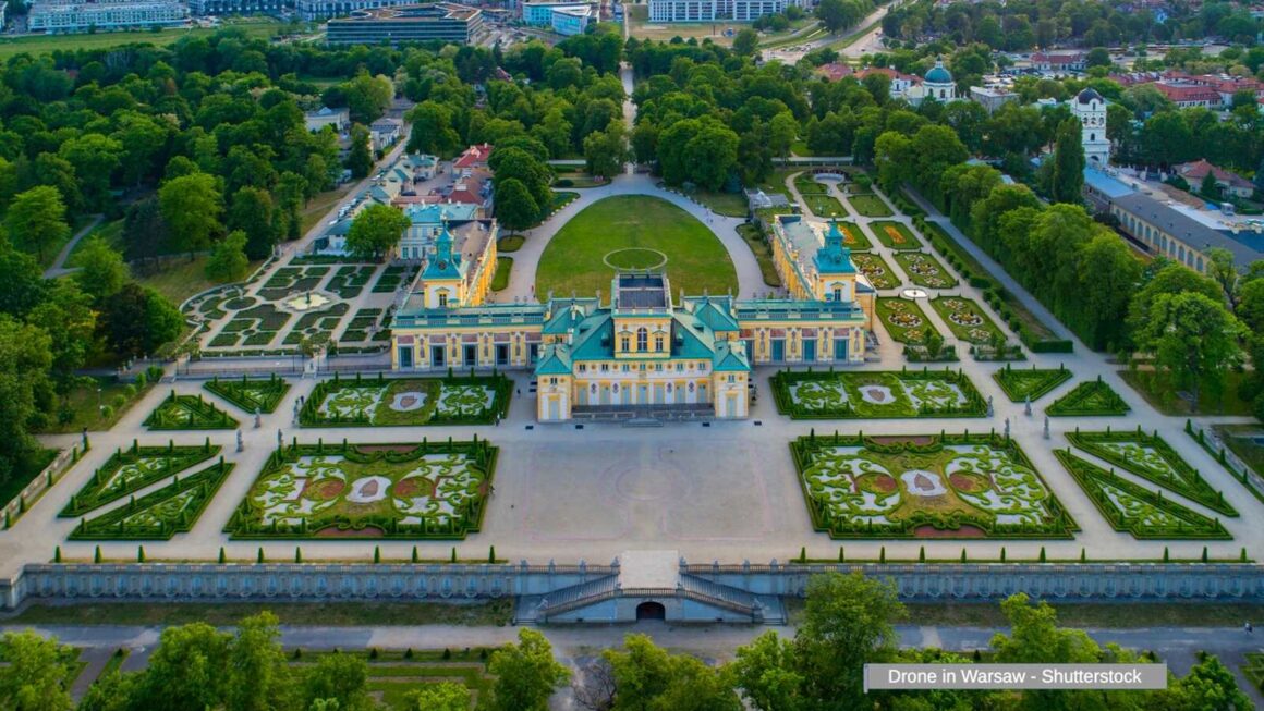
,Photo description: Top view in Wilanow – Royal Palace in Warsaw 05. October. 2018. Stock video production – Shutterstock.
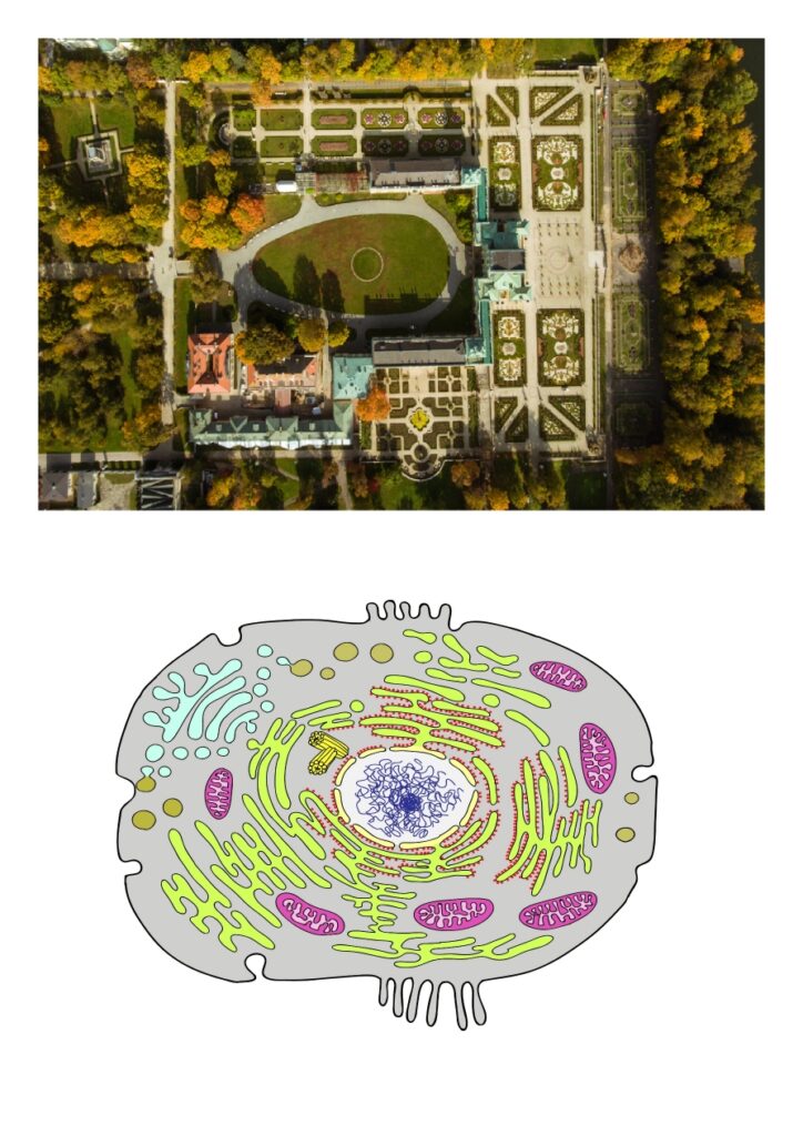
, Vector description: Animal cell, eukaryotic cell. Panaiotidi – Shutterstock.
The Architecture of Life
When viewed from above, the Wilanów Palace complex reveals a striking architectural blueprint that mirrors the fundamental structure of an animal cell. This is not mere coincidence—it appears to be an intentional encoding of cellular biology into baroque architecture.
The Nucleus – The Royal Heart
The central palace building, with its distinctive corps de logis and lateral wings, forms an irregularly shaped structure that corresponds to the cell nucleus. Just as the nucleus is not always perfectly spherical, the Wilanów palace complex extends asymmetrically, with the main building representing the nucleolus—the „command center within the command center”—while the side wings added by Elżbieta Sieniawska between 1723-1729 complete the nuclear architecture.
This is profound: the „royal palace” literally translates to „cell nucleus”—the residence of genetic royalty, the DNA that rules the cellular kingdom.
The Endoplasmic Reticulum – Gardens as Molecular Networks
Immediately surrounding the palace, the formal baroque gardens function as the endoplasmic reticulum—a network of membranes that in living cells extends directly from the nuclear envelope. Notice how these structured gardens flow seamlessly from the palace walls, just as the rough ER extends from the nuclear membrane in actual cells. This is not decorative coincidence; it’s architectural encoding of the secretory pathway.
The Golgi Apparatus – The Long Processing Center
In the northern section of the complex (lower left in aerial view), the longest building with its distinctive turquoise-green roof represents the Golgi apparatus—the cell’s processing and packaging center. Its elongated, flattened form perfectly mirrors the stacked cisternae structure of the actual Golgi apparatus in living cells. This building’s architecture encodes the Golgi’s function: receiving materials from one end (from the ER), processing them along its length, and packaging them for export at the other end.
The spatial organization is perfect: Nucleus (palace) → ER (immediate gardens) → Golgi (long turquoise building). The secretory pathway encoded in stone and gardens.
Supporting Structures – The Organelles
The other auxiliary buildings in the complex—including the building with the orange roof adjacent to the Golgi, along with the Kitchen Outbuilding and Guardhouse commissioned by Izabela Lubomirska—represent other organelles such as lysosomes and peroxisomes, the cell’s digestive and protective organelles. The pathways and alleys form the cytoskeleton, while the outer walls and boundaries of the estate define the cell membrane.
Functions of Structure of Animal Cells: Cells in most animals are categorised into higher levels of structures such as tissues, organs, and organ systems. Along with obtaining food and oxygen, animal cells also maintain their internal conditions, move and reproduce. A cell of an animal is highly specialised to accomplish a particular function. The organelles within each cell type are suited to the task that it performs.
Source: Unacademy – Structure of Animal CellWhat we’re witnessing is not simply a palace—it’s a three-dimensional textbook of cellular biology, written in stone, gardens, and baroque splendor by architects who possessed knowledge far beyond what conventional history attributes to the 17th and 18th centuries.
Video: WILANÓW PALACE │ WARSAW – Full walking tour by Lily around the world
„Sculptural Enzymes – The Molecular Workers”
Throughout the Wilanów gardens and integrated into the palace architecture, stone busts populate the landscape. At first glance, they appear to be classical decorative elements—portraits of emperors, philosophers, and nobles. But look closer at their three-dimensional form.
,Photo description: Gardens and flower beds in park of Wilanow Royal Palace in Warsaw, Poland.Liya_Blumesser – Shutterstock.
,Photo description: Rome, Italy-January 21, 2024: bust of the Roman Emperor Tiberius (Tiberius Julius Caesar Augustus). Dima Moroz – Shutterstock.
,Illustration description: Structure of ubiquitin carboxyl-terminal hydrolase isozyme L5 (Uch37) tetramer. 3D cartoon and Gaussian surface models, chain id color scheme, PDB 3ihr, white background. VD Image Lab – Shutterstock.
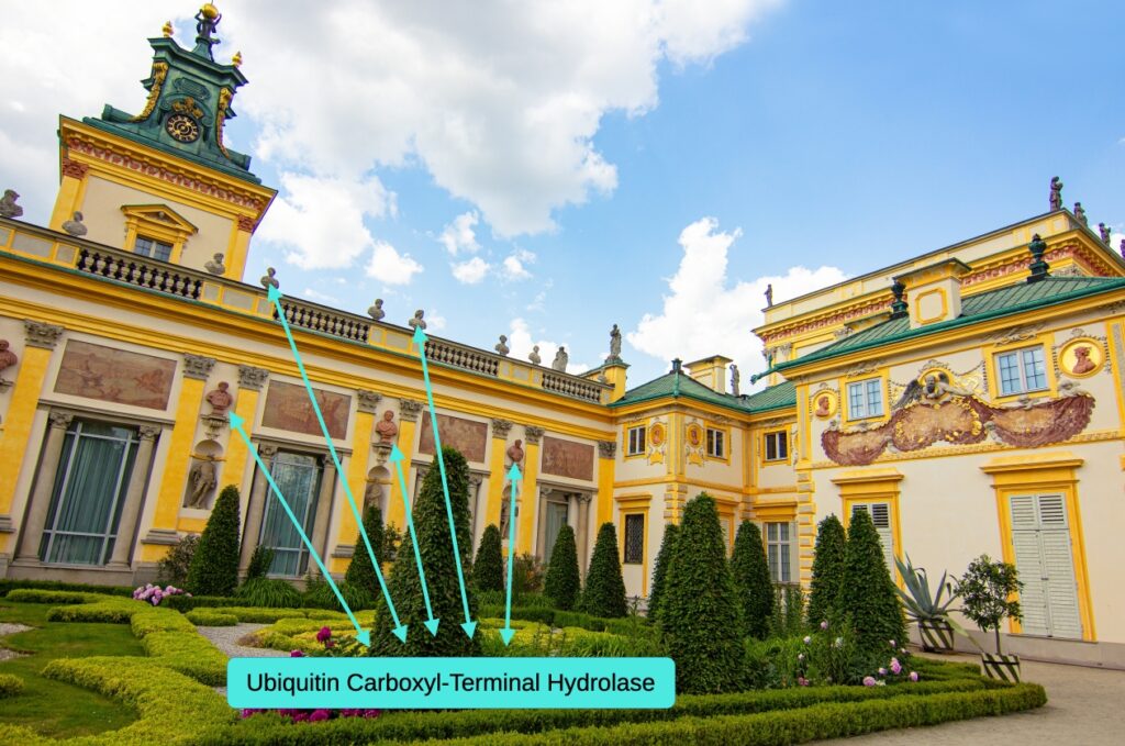
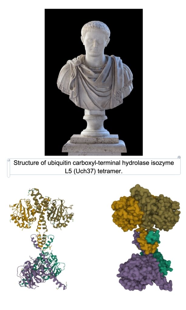
The external appearance of these sculptural busts mirrors the quaternary structure of complex enzymes—particularly the deubiquitinating enzymes that regulate protein degradation within cells.
Ubiquitin Carboxyl-Terminal Hydrolase 37 (UCH37, also called UCHL5) is a member of the deubiquitinating enzymes that can suppress protein degradation through disassembling polyubiquitin from the distal subunit of the chain. It has been proved that UCH37 can be activated by proteasome ubiquitin chain receptor Rpn13 and incorporation into the 19S complex. UCH37, which has been reported to assist in the mental development of mice, may play an important role in oncogenesis, tumor invasion and migration.
Source: Semantic Scholar – UCH37 ResearchUnfortunately, I cannot show the busts integrated into the Wilanów palace architecture up close in this post, so I’ve substituted them with another model from Rome. But observe: the external appearance of the bust sculpture is remarkably similar to the appearance of this enzyme structure.
The Diversity of Form
This enzyme belongs to the group of deubiquitinating enzymes, which means there are different types with different structures. It’s the same when you look at bust sculptures from all over the world—they have different shapes, different expressions, different profiles. Each unique form potentially encoding a different enzymatic structure.
See: Wikipedia – Bust (sculpture)There are many more figures around and on the royal palace. In my opinion, these are also enzyme structures involved in processes inside the cell, each sculpture a frozen moment of molecular machinery.
Every second inside every living cell, thousands of chemical reactions are taking place. These reactions constitute the essential tasks of life such as metabolism, protein synthesis, cell renewal and growth. The proteins called enzymes work to maintain the rate of these reactions at a life-sustaining level.
Video: How Enzymes Work by RCSBProteinDataBank
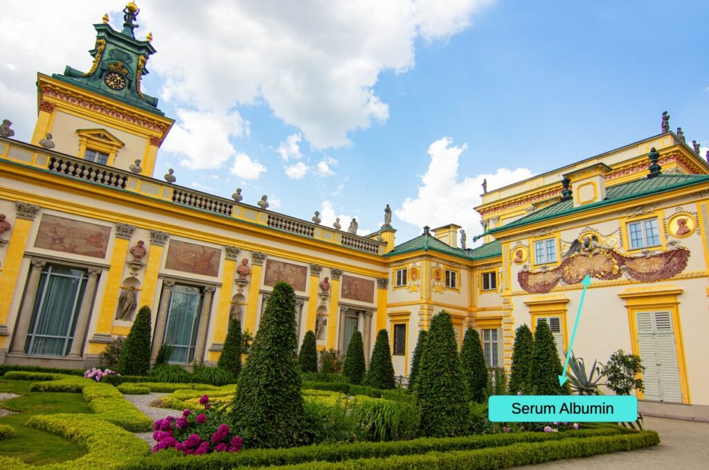
„The Sundial – Albumin’s Journey Through Time”
Time, Protein, and Transcytosis
On the garden façade of the south-eastern alcove stands one of Europe’s most beautiful sundials—a masterpiece that does far more than tell time. Commissioned by King John III Sobieski and drawn by the Jesuit Adam Adamandy Kochański (scientific adviser and librarian of the king), with architectural design by Augustin Locci, this is molecular biology disguised as baroque artistry.
,Photo description: Gardens and flower beds in park of Wilanow Royal Palace in Warsaw, Poland. Liya_Blumesser – Shutterstock.
,Photo description: Baroque solar clock on the wall of Wilanow castle. bykom photo – Shutterstock.
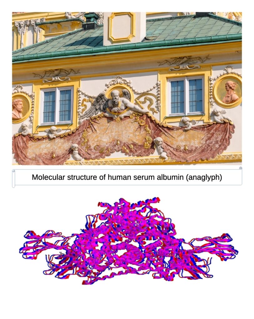
, Illustration description: Molecular structure of human serum albumin (anaglyph), example of globular protein, 3D rendering. Raimundo79 – Shutterstock.
The Composition:
The timepiece consists of three dials showing three types of clocks: Babylonian, local, and Italian. The gnomon on the central dial is held by winged Chronos, the god of time. On the side dials, the gnomon is held by putti—winged cupid figures.
Source: FotoPolska – Wilanów SundialThe Molecular Reading:
Serum albumin is a long-lived, highly abundant circulating protein. It binds water, circulating ions (K⁺, Na⁺), and fatty acids, and plays a crucial role in regulating oncotic pressure in the blood vessels. Albumin persistence is regulated by the neonatal crystallizable fragment receptor (FcRn), which binds it in a pH-dependent manner.
Source: The Jackson Laboratory – FcRn-Albumin InteractionsThe Encoded Mechanism:
The neonatal crystallizable fragment receptor (FcRn) facilitates the export of albumin by mediating transcytosis—the process of transporting macromolecules such as albumin through cells and ejecting them into the bloodstream.
Now observe the sundial composition:
- Chronos (winged, at top) → FcRn receptor – facilitates export, has „wings” for movement
- The Gnomon (the timepiece pointer) → Albumin serum protein – the object being transported
- Upper cupids (with wings) → Active transcytosis – mobile transport through cells
- Lower cupids (without wings) → Albumin stabilization – protection against degradation
Note carefully: The upper figures have wings and actively hold the gnomon, suggesting active transport. The lower cupids are wingless and hold the gnomon as if protecting it from degradation—they participate in the intermediate stages of transcytosis, providing albumin durability and helping transport it safely through cellular compartments.
Three types of time = Three phases of albumin transport
The three dials (Babylonian, local, Italian) may represent:
- Cellular uptake phase
- Transcytosis transport phase
- Blood vessel export phase
This is not a sundial. This is a diagram of protein trafficking, measuring not hours but the half-life of circulating albumin—which can persist in the bloodstream for 19-21 days, an extraordinarily long time for a protein. The „god of time” literally holds the long-lived protein.
The Pattern Emerges
We have now witnessed three levels of biological encoding in a single baroque palace:
- Cellular architecture – The whole complex maps to cell structure
- Enzymatic machinery – Sculptural busts encode enzyme quaternary structures
- Protein transport – The sundial diagrams albumin transcytosis
This is not coincidence. This is not pareidolia. This is systematic correspondence between sacred architecture and molecular biology.
The architects of Wilanów—Augustyn Locci, Adam Adamandy Kochański, Giovanni Spazzio—were not merely building a royal residence. They were encoding the fundamental mechanisms of life itself into stone, gardens, and baroque artistry. Every tower, every sculpture, every timepiece serves a dual purpose: aesthetic beauty and biological instruction.
What we call „decorative elements” are actually molecular diagrams. What historians dismiss as „symbolic ornamentation” is precise scientific documentation. The palace is simultaneously a home for a king and a textbook for those who know how to read it.
The question is no longer whether this encoding exists. The evidence is overwhelming. The question now is: Who taught them? And what other secrets remain hidden in the world’s sacred architecture, waiting for eyes that can see the molecular truth beneath the marble surface?
Combining images and analysis by Tomasz Mikulski – Cell God, date: 01/2025