🧬📚„The Map Was Always There. We Just Forgot How to Read It.”
For the past 10 months at CellGod.live, I’ve been documenting a pattern.
Churches that mirror NK cells. Fortresses replicating cellular defense systems. Parliamentary buildings organized like command centers. Sacred sites worldwide echoing the same structural logic as living organisms.
Each time, the same question surfaces: „How did ancient builders encode biological knowledge they shouldn’t have possessed?”
Today, I’m showing you where it all began. The source code. The original blueprint.
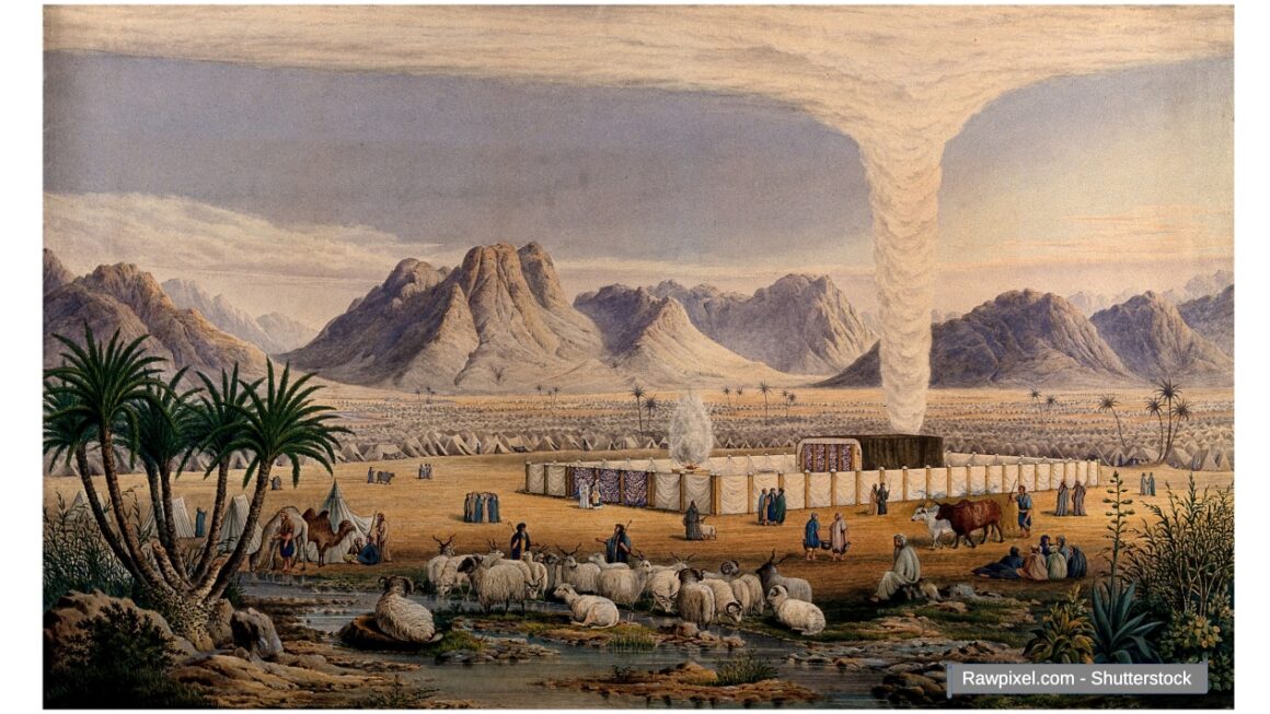
A Victorian illustration from 1850, based on a biblical text from 2,500-3,000 years ago.
Created by artist William Dickes, who thought he was simply drawing a religious camp in the desert.
But what he actually drew—without knowing it—was a complete map of a eukaryotic cell during active protein synthesis.
Not symbolically. Not metaphorically. Anatomically precise. Functionally accurate. Molecularly correct.
This illustration shows polyribosomes discovered in 1963, active chromatin loops understood in the 1970s, nuclear proteasomes mapped in the 1990s, and chromosome territories confirmed in the 2000s.
Every molecular structure was identified by science 103-140 years AFTER Dickes published his illustration. Every structure was described in the biblical text 2,500-3,000 years BEFORE cells were discovered.
This isn’t another „church looks like a cell” observation. This is the Rosetta Stone.
If the biblical Tabernacle is actually a cellular diagram encoded in religious language, then every temple, church, and sacred structure built on those principles becomes an echo of that original code. The builders weren’t creating art. They were transmitting information.
And here’s what changes everything.
The text identifies „the chosen people” as the Israelites moving through this architectural space. But in the cell, the molecules „chosen by DNA” to translate genetic code are ribosomes. Not a nation. Not an ethnicity. Molecular machines.
Every human has the same „chosen” ribosomes. A Jew has 80S ribosomes. An Arab has 80S ribosomes. You have 80S ribosomes. Identical. Universal. Equal.
If „chosen people” is truly a biological metaphor for ribosomal function, then millennia of religious conflict and ethnic supremacy have been fought over a fundamental misunderstanding. A molecular process mistaken for divine ethnic selection.
„Today, you’ll see how every major element of the Tabernacle—from the rising smoke to the corner tent—maps onto cellular architecture with remarkable precision. Spatial relationships matching modern cell biology. Functional analogies discovered decades or even a century after the illustration was published.”
You’ll see smoke rising as active transcription. Altars performing quality control. Gates functioning as nuclear pores. The „baptism of ribosomes” in nucleoplasm.
And you’ll confront the question that haunts every discovery on this blog: How did they know?
Welcome back to CellGod.live, where ancient architecture meets molecular biology, and where the line between „sacred text” and „scientific blueprint” disappears entirely.
Let’s decode the Tabernacle.
– Illustration description: Historical illustration depicting the Tabernacle in the wilderness, surrounded by tents and people, showing an ancient religious scene with a central sacred structure. Religious illustration from the period, work of art. (RawPixel.com – Shutterstock)
– Illustration description: Biology Illustration of the Animal cell, (Art Garibalda – Shutterstock)
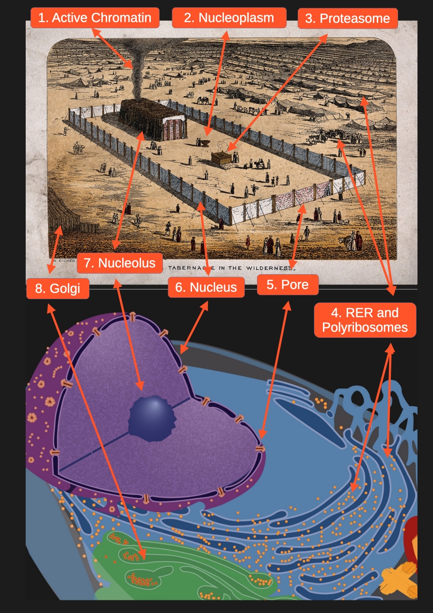
Let’s analyze the illustration element by element. Each component has its molecular counterpart.
- 1. Active Chromatin: Smoke as the Genome in Action
The smoke emerging from the tent and forming a cloud above the courtyard is probably the most ingenious, unconscious symbol. Its swirling, thread-like forms represent active euchromatin – chromosomal loops carrying rRNA genes that extend into the nucleolus during transcription. The cloud above the courtyard beautifully represents chromosome territories – distinct spatial regions where individual chromosomes organize themselves within the nuclear space.
What we see in the Tabernacle: Swirling smoke emerging from the central tent, rising to form a cloud above the courtyard. Thread-like, dynamic forms creating a canopy over the sacred space.
What it really is: Active euchromatin – specifically nucleolar organizing regions (NORs) with rRNA genes looping into the nucleolus, plus chromosome territories filling the nuclear space like clouds in distinct atmospheric zones.
Why this metaphor is perfect: Thread-like forms mirror DNA’s structure. The swirling represents chromatin opening during transcription. Emergence from the hidden center makes invisible genetic activity visible. The cloud above represents chromosome territories – each chromosome occupying its own spatial domain within the nucleus, like clouds filling the sky without mixing.
The molecular reality: The nucleolus forms around actively transcribing rRNA genes through liquid-liquid phase separation. Chromatin loops enter this dense core for transcription. Meanwhile, chromosome territories organize throughout the nucleus – each chromosome in its distinct domain, maintaining genomic order in three-dimensional space.
Biblical context: The cloud covered the Tabernacle as divine presence and guidance – dynamic, not static.
Molecular context: Active transcription is the cell’s „pillar of cloud” – genetic activity made manifest. The smoke rising into territorial clouds captures both local transcription and global chromosomal organization.
Note: The cell diagram doesn’t show these chromatin dynamics or chromosome territories – they’re visible only under specialized microscopy as distinct regions within the nucleus. - 2. Nucleoplasm: The Basin as Nuclear Medium
The basin (or container) of water symbolizes the nucleoplasm – the dense, fluid matrix filling the interior of the nucleus, in which all nuclear structures are immersed. The two figures beside it are newly synthesized ribosomal subunits that have left the nucleolus.
What we see in the Tabernacle: A large container/basin with water. Two figures standing next to it.
What it really is: Nucleoplasm – dense, fluid matrix filling the interior of the nucleus.
Most beautiful analogy: „Baptism of Ribosomes”
What happens: Ribosomes are synthesized in the nucleolus (Holy of Holies), exit as two separate subunits (large and small), „immerse” in the nucleoplasm, undergo quality control, are exported through nuclear pores to the cytoplasm, where they join into a functional machine.
Two figures in the basin = two ribosomal subunits: Large subunit (60S), Small subunit (40S).
„Baptism” means: Preparation, purification, consecration before mission, transformation (from unready to ready).
Note: The cell diagram shows nucleoplasm as the interior space of the nucleus, though molecular machines like assembling ribosomal subunits are too small to be visible at this scale. - 3.Nuclear Quality Control: Altar as Nuclear Proteasome
The altar of burnt offering, positioned within the courtyard (representing the nucleus itself, not the cytoplasm), represents nuclear proteasomes – the quality control system that ensures only properly assembled ribosomes and functional proteins leave the nucleus.
What we see in the Tabernacle: An altar inside the courtyard, where animals (cattle, sheep) are brought for sacrifice. Human figures with livestock standing near the altar, awaiting inspection. Individual human figures scattered throughout the courtyard represent maturing ribosomal subunits undergoing final assembly and quality control before export.
What it really is: Nuclear proteasomes performing quality control – the cellular „inspection station” where proteins are checked for defects before they can function or be exported.
Why this spatial location is perfect: The altar is inside the courtyard (= inside the nucleus), positioned before the gate (= before nuclear pores). Animals must pass inspection before leaving (= proteins checked before export to cytoplasm).
The biblical process: Animals brought to altar → Priests inspect for defects → Defective animals „burned” (sacrificed) → Ashes recycled as fertilizer → Only „pure” animals accepted.
The cellular process: Proteins enter nucleus → Nuclear proteasomes „inspect” them → Defective proteins ubiquitinated → Proteasomes degrade them to amino acids (recycling) → Only properly functioning proteins complete their mission.
The livestock near the altar represent transcription factors and regulatory proteins – molecular messengers that enter the nucleus from cytoplasm to perform specific tasks (regulating gene expression, controlling cell cycle), then are degraded by nuclear proteasomes after completing their mission. Like sacrificial animals brought from camp into sacred courtyard to fulfill their purpose then consumed on altar, these proteins journey from cytoplasm to nucleus, execute their function, and are destroyed when no longer needed.
Scientific fact: Transcription factors like p53, NF-κB, and c-Myc are synthesized in cytoplasm, imported into nucleus to regulate DNA transcription, then rapidly degraded by nuclear proteasomes. Additionally, approximately 20-30% of newly imported ribosomal proteins are immediately degraded by nuclear proteasomes through Nuclear Ribosome Quality Control (NRQC). About 40% of all proteasomes in a cell are located in the nucleus.
Theological parallel: In Judaism, sacrifice is transformation – converting impure to pure. Similarly, the nuclear proteasome transforms potentially harmful proteins into useful amino acids, ensuring nothing defective enters the cytoplasm.
Key insight: The courtyard isn’t the cytoplasm—it’s the nucleus itself. The individual figures scattered throughout represent maturing ribosomal subunits preparing for export. The altar isn’t outside the cell—it’s inside the nucleus, performing critical quality control to ensure only functional proteins complete their nuclear mission. - 4. RER and Polyribosomes: Camp Organization as Protein Factories
The vast desert is the cytoplasm – the cell’s basic matrix. The orderly rows of camp tents are sites of the rough endoplasmic reticulum (RER), the main location of exported protein synthesis. The Israelites gathered in small clusters throughout the camp and desert alongside their livestock represent polyribosomes – groups of ribosomes working together on the same mRNA strand, creating multiple copies of the same protein simultaneously.
What we see: A huge desert space surrounding the central camp, orderly rows of tents, and groups of Israelites with their livestock.
What it really is: Cytoplasm (dense gel-like matrix filling 70% of cell’s interior), RER (endoplasmic reticulum covered with ribosomes), and polyribosomes (multiple ribosomes on single mRNA strand).
Why it fits: The desert teems with life like cytoplasm. The organized tent rows surround the center like RER surrounds the nucleus. The clustered people and animals mirror polyribosome structure under electron microscopy.
The people (Israelites) represent ribosomes – the molecular machines chosen to translate mRNA. The livestock represent proteins being synthesized – including transcription factors and regulatory proteins destined for the nucleus, and structural proteins remaining in cytoplasm. Some animals (proteins) will journey through the gate into the courtyard (nucleus) to perform regulatory functions before being sacrificed at the altar (degraded by proteasomes).
Scientific fact: Under electron microscopy, polyribosomes appear as clusters of dark dots (ribosomes) connected by thin thread (mRNA), with fuzzy extensions (growing proteins) emerging from each ribosome – exactly like groups of people with livestock in formation. Linear arrangements along mRNA resemble people and animals in procession, with groups varying in size (3-10 ribosomes per mRNA, like families with herds).
Poetic truth: The illustration shows molecular reality of protein synthesis as a pastoral scene of shepherds with flocks. Each „herd” represents proteins being simultaneously produced, some destined for the sacred journey into the nucleus. - 5. Nuclear Pore Complex: Gate as Selective Gateway
The gate leading to the courtyard is literally the nuclear pore complex – a selective, guarded gate controlling all exchange between the interior of the nucleus and the cytoplasm.
What we see in the Tabernacle: A gate leading to the courtyard, guarded and controlled. Individual figures scattered throughout the courtyard (maturing ribosomal subunits) will exit through this gate once properly assembled.
What it really is: Nuclear Pore Complex (NPC) – the most advanced „gate” in biology. 30 different types of proteins (nucleoporins) selectively pass molecules, „check passes” (nuclear localization signals), and actively transport.
Similarity: Like the biblical gate guarded by Levites controlling who enters the holy place, the nuclear pore allows only molecules with correct „molecular passport” (nuclear localization signals) to enter the nucleus. Transcription factors synthesized in cytoplasm must present correct signals to pass through. Ribosomal subunits assembled in nucleolus must be properly formed to exit. The gate perfectly discriminates – allowing inward traffic of regulatory proteins and outward traffic of ribosomes.
Biblical context: The gate separated sacred courtyard (where God dwelt) from common camp. Levites stood guard, checking credentials. Only ritually pure could enter.
Molecular context: The nuclear pore separates nucleus (where genetic information is stored) from cytoplasm. Nucleoporins act as guards, checking „credentials” (signal sequences). Only authorized molecules can pass – transcription factors going in, ribosomes coming out.
In the cell diagram: The small holes in nuclear envelope represent these nuclear pores – checkpoints between nucleus and cytoplasm, the same gates through which transcription factors enter to regulate genes and „baptized” ribosomal subunits (the individual figures from the courtyard) exit to begin their mission in the cytoplasm, where they will join into functional ribosomes. - 6. Nucleus: The Tabernacle Courtyard as Command Center
The courtyard fence constitutes the nuclear envelope – a double membrane barrier separating the sacred sanctuary of the nucleus from the rest of the cell. The pillars supporting the fence are the nuclear lamina – a network of proteins providing the nucleus with mechanical strength and serving as an anchor point for chromatin.
What we see in the Tabernacle: A fence surrounding the tabernacle, creating a sacred courtyard. Pillars supporting the structure. The boundary between the holy space and the common camp.
What it really is: Nuclear Envelope (double membrane) and Nuclear Lamina (structural support).
Fence = Nuclear Envelope: In the illustration we see a single fence representing the nuclear boundary. In reality, the nuclear membrane is double-layered – an outer membrane continuous with the RER, and an inner membrane studded with lamina proteins. The biblical description may have simplified this to a single fence, or the artist chose to represent both membranes as one structure. The function remains the same – it separates the sacred (nucleus) from the secular (cytoplasm), protects the genetic information within, and is continuous with the RER (like a fence connecting to the camp structures).
Pillars = Nuclear Lamina: Network of proteins (lamins A, B, C), provides mechanical stability, anchor points for chromatin, maintains nuclear shape and integrity. These are the structural „pillars” that prevent nuclear collapse.
Scientific fact: Mutations in lamin genes cause progeria – premature aging disease. When the structural pillars fail, the entire „sanctuary” collapses. This shows how CRUCIAL these support structures are – just as the biblical pillars held up the courtyard fence.
Symbolic parallel: The rectangular courtyard in the Tabernacle represents the functional architecture of the nucleus – not its literal spherical shape, but its organizational logic. Like a circuit diagram versus an actual circuit, or like the London Underground map versus actual winding tracks, it shows relationships rather than physical form. The Bible describes an idealized, geometrically perfect sanctuary because it’s encoding functional principles, not literal shapes.
In the cell diagram: The circular boundary clearly shows the nuclear envelope surrounding everything within – the nucleolus, nucleoplasm, and all nuclear machinery. Though in the Tabernacle it’s depicted as rectangular, this represents the same functional boundary – the sacred space where genetic information is protected and processed. - 7. Nucleolus: The Holy of Holies as Ribosome Factory
The central Tent of the Tabernacle represents the nucleolus—the ribosome factory within the nucleus, the most protected structure where the machinery for building ribosomes is housed. The nucleolus contains multiple zones with different functions, and the innermost chamber—the Holy of Holies—corresponds to the Dense Fibrillar Component (DFC), the zone where ribosomal DNA is actively transcribed. This is the most sacred, most protected space within the nucleolus—the place where genetic silence becomes genetic voice, where DNA is read and RNA emerges. What we see in the Tabernacle: The largest, most protected structure at the center of the camp, surrounded by a courtyard and veils. The innermost sanctuary, accessible only to the high priest once per year. What it really is: The nucleolus within the cell nucleus—the ribosome factory, where rRNA is transcribed (in the DFC/Holy of Holies) and ribosomal subunits are assembled (in the surrounding zones). Why it fits: Central location within the nucleus, greatest protection (nested within the nucleus itself), controlled access, contains the machinery for rRNA transcription and ribosome assembly, occupies about 10% of nuclear space—just as the Holy of Holies occupied about 10% of the Tabernacle structure. Key observation: The Bible describes the Holy of Holies as inaccessible to most people—only the high priest once a year, and even then only after elaborate purification rituals. Similarly, most molecules do NOT have access to the transcriptional core of the nucleolus—only selected proteins involved in ribosome biogenesis can enter this sacred space. The nucleolus maintains its boundaries through liquid-liquid phase separation—a molecular form of „sacred space” maintained by specific protein interactions. Biblical parallel: The Holy of Holies contained the Ark of the Covenant with the tablets of the Law—the fundamental instructions for the nation. The nucleolus contains rDNA—the genes encoding ribosomal RNA, the fundamental instructions for protein synthesis in every living cell. We’ll explore what the Ark itself represents in Part II. In the cell diagram: The dark sphere within the nucleus clearly shows the nucleolus—the „tent” of the cell, where the sacred work of ribosome creation occurs continuously. - 8. Golgi Apparatus: Corner Tent as Sorting Center
The tent protruding in the lower left corner, with characteristic layered „stripes” (rolled fabrics or skins), is strikingly similar to the stacks of cisternae in the Golgi apparatus. This is the center for sorting, labeling, and packaging cellular products.
What we see in the Tabernacle: A tent in the lower corner with characteristic layered „stripes” – rolled fabrics or skins stacked in parallel layers.
What it really is: Golgi apparatus – center for sorting, packaging, and modifying proteins before they’re sent to their final destinations.
Why it looks strikingly similar: Golgi apparatus under microscope – Stack of flattened, layered cisternae, looks like „stacked pancakes,” 5-8 layers arranged in parallel. Tent in illustration – Layered strips of material, folded, parallel lines, clearly separated from other structures.
Function: Proteins come from RER (camp tents), are modified with sugar chains (glycosylation), sorted by destination, packaged into vesicles, receive „molecular addresses” (targeting signals indicating where they should go), sent in transport vesicles to target destinations (cell membrane, lysosomes, or secretion outside the cell).
Human figures between tent and Tabernacle = proteins in transport. These represent the constant flow of newly synthesized proteins moving from the RER through the Golgi, receiving their modifications and sorting signals before being shipped to their final locations.
Biblical parallel: Just as the Tabernacle had a system for sorting offerings and sacred objects – some remained in the sanctuary, some were distributed to priests, some were sent out to the tribes – the Golgi sorts proteins into different categories based on their ultimate purpose.
In the cell diagram: The layered structure in the cytoplasm clearly shows the Golgi apparatus – matching the layered tent in the Tabernacle illustration. It sits between the RER and the cell membrane, perfectly positioned for its role as the cell’s „post office” – receiving, processing, and shipping proteins to their destinations.
🔥 KEY DISCOVERY: Israelites = RIBOSOMES
This is not a random analogy. This is the key to understanding the greatest perversion in human history.
Think about it:
The biblical „people chosen by God” are ribosomes chosen by DNA to translate the universal genetic code.
NOT a specific nation of people. NOT Jews, Arabs, or anyone else. MOLECULES. CELLULAR MACHINES.
Why Are Ribosomes „Chosen”?
Among thousands of types of molecules in the cell, only ribosomes have the privilege of reading the sacred code (mRNA), translating genetic instructions, creating proteins – the foundation of life, direct contact with information from DNA.
Every human has the same „chosen” ribosomes: A Jew has 80S ribosomes. An Arab has 80S ribosomes. You have 80S ribosomes. I have 80S ribosomes.
Identical. Universal. Equal.
Consequences of This Discovery:
If „chosen people” is a metaphor for ribosomal function, not human superiority, then every human carries within quintillions of „chosen ones” (ribosomes), chosenness is molecular, not ethnic, we are all equally „sacred” at the biological level, using this metaphor for supremacy = sacrilege.
But more on this in the next post. Now let’s return to the analysis…
🎯 Why Is the Nucleus Rectangular? (Symbol vs Reality)
An attentive reader will ask: „But cell nuclei are round! Why is the Tabernacle rectangular?”
Excellent question.
Dickes faithfully recreated the biblical description, which uses ideal geometry as a symbol of divine order.
In nature, nuclei are usually spherical, but can be elongated, irregular, shape depends on cell type.
The rectangle is a symbolic architectural plan, ideal functional scheme, cell „logo” (like electrical diagram vs actual circuit).
In biology we call this „cell logic” – a simplified model showing functional relationships, not literal shape.
Analogy: The London Underground map shows straight lines between stations. The actual tracks are winding. But the map is more useful than a literal plan.
Similarly, the Tabernacle shows functional cell logic, not the literal appearance under a microscope.
🧩 What’s Missing? (Undeciphered Organelles)
An attentive observer will notice: not all organelles are in the illustration.
Missing are mitochondria (energy powerhouses), cytoskeleton (structural framework), peroxisomes (detoxification), lysosomes (digestion), smooth endoplasmic reticulum (lipid synthesis).
Does this mean the theory is wrong?
No. There are several explanations:
1. The illustration shows a cell in interphase – a state of active transcription and translation
Dickes painted a close-up of the main organelles involved in the Central Dogma of Molecular Biology: DNA → RNA → Protein (Ark → Smoke → Ribosomes).
It’s like a microscopic view focused on the nucleus, RER, and cytoplasm during intense protein synthesis. Other organelles are outside the frame or in the background.
2. Dickes deciphered what the biblical text described
The Book of Exodus (chapters 25-40) describes in detail the Tabernacle, its interior, altars, and camp layout – i.e., structures related to:
- Information storage (Ark = DNA)
- Production (Tent = ribosome synthesis)
- Translation (Israelites = ribosomes/polyribosomes)
Other organelles may be encoded elsewhere.
Examples of potential encodings:
Cytoskeleton: Detailed dimensions of tent ropes, systems of stakes and supports in descriptions of the Tabernacle’s construction.
Smooth endoplasmic reticulum: Mountains on the title illustration.
Peroxisomes and lysosomes: Purification rituals and sacrifice „digestion” processes may encode these degradative organelles.
This remains to be discovered in other biblical texts and contexts.
Summary: Dickes didn’t draw a complete cell – he drew a moment in the cell’s life: interphase with active protein synthesis. It’s like a photograph focused on one process, not a complete cytology atlas.
📊 Summary – The Macroscale of Life
Let’s see the full picture of what Dickes unknowingly drew:
Dickes’ Illustration = Cell in Interphase
Cell state: Intense transcription and translation
Complete analogy table:
Desert = Cytoplasm (Basic matrix), Israelite groups = Polyribosomes (Coordinated protein synthesis), Camp tents = RER (Exported protein synthesis), Central Tent = Nucleus (DNA protection), Interior of tent = Nucleolus (Ribosome production), Smoke = Euchromatin (Active transcription), Gate = Nuclear pores (Transport control), Fence = Nuclear membrane (Protective barrier), Pillars = Nuclear lamina (Structural stability), Altar = Proteasome (Protein degradation), Basin = Nucleoplasm (Nuclear medium), Corner tent = Golgi apparatus (Protein sorting)
This is not a metaphor. This is a MAP.
🔮 What Does This All Mean?
A Pattern Across Millennia
William Dickes’ 1850 illustration isn’t an isolated curiosity. It’s one data point in a pattern that spans continents and centuries.
But before we explore that pattern, consider what makes this discovery so extraordinary:
Timeline of Scientific Discovery:
1850 – Dickes publishes his illustration based on a text written 2,500-3,000 years earlier
Scientists in 1850 understood that cells had nuclei (discovered 1831-1833). They knew cells existed. That was essentially it.
Then came the discoveries that revealed what Dickes had unknowingly drawn:
1953 (103 years later) – Watson & Crick discover DNA’s double helix structure
1955 (105 years later) – Scientists understand how ribosomes synthesize proteins
1963 (113 years later) – Polyribosomes discovered – multiple ribosomes working coordinately on the same mRNA strand, exactly as depicted in the clusters of Israelites with their livestock
1970s (120+ years later) – Chromatin dynamics during transcription finally understood – the mechanism by which DNA loops extend into the nucleolus for rRNA synthesis, perfectly matching the rising smoke from the sanctuary
1990s (140+ years later) – Nuclear ribosome quality control pathway discovered – the system by which defective ribosomal proteins are degraded by nuclear proteasomes before export, precisely corresponding to the altar with sacrificial animals inside the courtyard
Every molecular detail Dickes drew existed in the ancient biblical text long before microscopes could see it. Existed in his 1850 illustration long before scientists could name it. And exists in our cells right now, functioning exactly as the ancient description specified.
The Pattern Repeats Across Millennia:
Timeline:
- ~1500-1000 BCE: The Book of Exodus describes the Tabernacle
- ~500 BCE: Kalai-Tepe fortress (Tajikistan) – architectural cell model, https://cellgod.live/tajikistan-fortress-cup-kalai-teppe-in-istaravshan/
- 1835-1839: Scientists formally describe the nucleolus
- 1850: Dickes creates his illustration from ancient text
- 1878: Chromosomes are named
- 1953: DNA structure discovered
- 1955: Ribosome function understood
- 1963: Polyribosomes discovered
- 1970s: Chromatin dynamics understood
- 1990s: Nuclear quality control discovered
- 2025: We notice the complete pattern
The pattern appears in: Ancient texts (Torah, 2,500-3,000 years old), Medieval architecture (fortresses, temples), Baroque churches (NK cell structures), Parliamentary buildings ( command centers), Sacred sites worldwide (cellular organization)
Three Possible Explanations:
1. Extraordinary Coincidence
The probability that this architectural elements align with cellular structures—with correct spatial relationships and functional analogies—by pure chance?
Consider what would need to align by accident:
- Polyribosomes (discovered 1963) matching clusters of people with livestock
- Active chromatin loops (understood 1970s) matching rising smoke
- Nuclear proteasomes (discovered 1990s) matching the altar’s location and function
- Nucleoplasm matching the basin where ribosomal subunits undergo final preparation
- All positioned correctly in relation to each other
The probability is astronomically low. Like flipping heads 50 times in a row.
2. Universal Principles of Organization
Perhaps all complex systems—biological, architectural, social—naturally evolve similar organizational patterns:
- A protected center (nucleus/sanctuary)
- Production zones (ER/workshops)
- Transport networks (cytoskeleton/corridors)
- Waste processing (proteasomes/altars)
- Quality control checkpoints (nuclear pores/gates)
- Controlled borders (membranes/walls)
This wouldn’t require ancient knowledge of cells—just intuitive recognition of what makes complex systems work efficiently.
But this explanation struggles with the specificity: Why do the exact molecular processes discovered in the 1960s-1990s appear in a text from 2,500-3,000 years ago? Universal principles might explain general similarities, but not precise details like polyribosomes, chromatin dynamics, and nuclear quality control.
3. Unknown Knowledge Transmission
The most controversial explanation: The ancients possessed observational or conceptual tools we don’t understand.
This doesn’t require believing in lost technologies or supernatural revelation—only acknowledging that:
- Human understanding isn’t linear
- Knowledge can be encoded in forms that survive millennia
- What appears as „religious symbolism” might preserve information in ways we’re only now learning to decode
The challenge: If this is true, how did they know? What method of observation or transmission allowed knowledge of molecular processes to be encoded in architectural descriptions?
The Question That Remains:
We can debate how this knowledge was encoded. But the fact remains undeniable:
A text written 2,500-3,000 years ago describes a structure that maps onto eukaryotic cell organization with stunning precision—including molecular details that science didn’t discover until 103-140 years after Dickes drew his illustration.
An artist in 1850, faithfully illustrating that ancient text, created what we now recognize as cellular architecture at a molecular level he couldn’t possibly have understood.
And sacred buildings worldwide echo these same patterns.
Is this design? Intuition? Coincidence? Or something we haven’t yet named?
The answer matters less than the question it forces us to ask:
What else might be encoded in plain sight, waiting for us to learn how to see it?
And if the concept of „chosen people” is actually a metaphor for ribosomes—the molecular machines chosen by DNA to translate the code of life—what does this mean for millennia of religious and ethnic conflict built on misunderstanding this metaphor?
🚀 What’s Next?
What you’ve seen today is just the surface.
We’ve looked at the Tabernacle from the outside – from the perspective of the desert. But what lies behind the veil?
The Holy of Holies holds secrets we haven’t yet touched upon.
The Ark of the Covenant contains three sacred objects. Ancient texts describe them with precision that seems… suspiciously specific. As if someone wanted to preserve information in a form that would survive millennia.
In the next post, we’ll step inside.
You’ll see a three-dimensional reconstruction of the interior. And you’ll discover why the arrangement of these objects, their materials, and their symbolism form something far more profound than religious metaphor.
We’ll answer the question that changes everything:
If the „chosen people” is truly a metaphor for ribosomal function… what does this mean for how we understand identity, purpose, and the sacred texts that have shaped civilizations?
The veil is about to be lifted.
Are you ready to see what our ancestors encoded in the heart of the sanctuary?
💭 Before You Dive Deeper… Take a Moment.
This discovery isn’t just about ancient texts or biology. It’s about the foundations of how you perceive the world.
- Which of these parallel stories resonated most strongly with your reason or your heart?
- Is there something in your own knowledge or beliefs that echoes this vision?
- For you – what is this? The strangest coincidence in history? The voice of the human soul? Or perhaps… a sign?
Your perspective is unique and essential. Leave a trace. Share this with someone who would be intrigued. Truth, on any level, demands to be spoken.
In the next part, I invite you inside:
Next post: „⚗️ Behind the Veil – How the Ark of the Covenant Contains the Instruction Manual for Building Ribosomes”
Combining images and analysis by Tomasz Mikulski – Cell God, date: 10/2025
- 📚 SOURCES, REFERENCES & FURTHER READING
Scientific Literature on Nucleolar Structure and Function
Sub-nuclear compartments
https://www.researchgate.net/figure/Sub-nuclear-compartments-Reproduced-with-the-permission-from-211_fig1_275336681
„The Nucleolus: Core of Ribosomal Mastery”
https://youtube.com/shorts/JSaCj8avF9k?si=7PO6UIJSSeroSLAm
Pederson, T. (2011)
„The nucleolus.” Cold Spring Harbor Perspectives in Biology, 3(3), a000638.
https://doi.org/10.1101/cshperspect.a000638
https://cshperspectives.cshlp.org/content/3/3/a000638
Boisvert, F.M., van Koningsbruggen, S., Navascués, J., & Lamond, A.I. (2007)
„The multifunctional nucleolus.” Nature Reviews Molecular Cell Biology, 8(7), 574-585.
https://doi.org/10.1038/nrm2184
Németh, A., & Längst, G. (2011)
„Genome organization in and around the nucleolus.” Trends in Genetics, 27(4), 149-156.
https://doi.org/10.1016/j.tig.2011.01.002
Ribosome Biogenesis
Henras, A.K., Plisson-Chastang, C., O’Donohue, M.F., Chakraborty, A., & Gleizes, P.E. (2015)
„An overview of pre-ribosomal RNA processing in eukaryotes.” Wiley Interdisciplinary Reviews: RNA, 6(2), 225-242.
https://doi.org/10.1002/wrna.1269
Woolford, J.L., & Baserga, S.J. (2013)
„Ribosome biogenesis in the yeast Saccharomyces cerevisiae.” Genetics, 195(3), 643-681.
https://doi.org/10.1534/genetics.113.153197
Ribosomal Protein Synthesis and Import
Warner, J.R. (1999)
„The economics of ribosome biosynthesis in yeast.” Trends in Biochemical Sciences, 24(11), 437-440.
https://doi.org/10.1016/S0968-0004(99)01460-7
Key finding: Ribosomal proteins are synthesized in the cytoplasm and imported into the nucleus.
Kressler, D., Hurt, E., & Baßler, J. (2010)
„Driving ribosome assembly.” Biochimica et Biophysica Acta, 1803(6), 673-683.
https://doi.org/10.1016/j.bbamcr.2009.10.009
Details the complete pathway from cytoplasmic synthesis to nuclear assembly.
Nuclear Ribosome Quality Control (NRQC)
Sung, M.K., Porras-Yakushi, T.R., Reitsma, J.M., Huber, F.M., Sweredoski, M.J., Hoelz, A., Hess, S., & Deshaies, R.J. (2016)
„A conserved quality-control pathway that mediates degradation of unassembled ribosomal proteins.” eLife, 5, e19105.
https://doi.org/10.7554/eLife.19105
Demonstrates that 20-30% of newly synthesized ribosomal proteins are degraded by nuclear proteasomes.
Defenouillère, Q., Yao, Y., Mouaikel, J., Namane, A., Galopier, A., Decourty, L., Doyen, A., Malabat, C., Saveanu, C., Jacquier, A., & Fromont-Racine, M. (2013)
„Cdc48-associated complex bound to 60S particles is required for the clearance of aberrant translation products.” Proceedings of the National Academy of Sciences, 110(13), 5046-5051.
https://doi.org/10.1073/pnas.1221724110
Quality control mechanisms for ribosomal proteins before nucleolar entry.
Polyribosomes (Polysomes)
Warner, J.R., Knopf, P.M., & Rich, A. (1963)
„A multiple ribosomal structure in protein synthesis.” Proceedings of the National Academy of Sciences, 49(1), 122-129.
https://doi.org/10.1073/pnas.49.1.122
Original discovery of polyribosomes – multiple ribosomes on single mRNA.
Brandt, F., Etchells, S.A., Ortiz, J.O., Elcock, A.H., Hartl, F.U., & Baumeister, W. (2009)
„The native 3D organization of bacterial polysomes.” Cell, 136(2), 261-271.
https://doi.org/10.1016/j.cell.2008.11.016
Structural analysis of polyribosome organization.
rDNA and Nucleolar Organizing Regions (NORs)
McStay, B., & Grummt, I. (2008)
„The epigenetics of rRNA genes: from molecular to chromosome biology.” Annual Review of Cell and Developmental Biology, 24, 131-157.
https://doi.org/10.1146/annurev.cellbio.24.110707.175259
Tchurikov, N.A., Fedoseeva, D.M., Sosin, D.V., & Snezhkina, A.V. (2015)
„Hot spots of DNA double-strand breaks in human rDNA units are produced in vivo.” Scientific Reports, 5, 10866.
https://doi.org/10.1038/srep10866
Nuclear Bodies and Compartmentalization
Mao, Y.S., Zhang, B., & Spector, D.L. (2011)
„Biogenesis and function of nuclear bodies.” Trends in Genetics, 27(8), 295-306.
https://doi.org/10.1016/j.tig.2011.05.006
Sawyer, I.A., Sturgill, D., & Dundr, M. (2019)
„Membraneless nuclear organelles and the search for phases within phases.” Wiley Interdisciplinary Reviews: RNA, 10(2), e1514.
https://doi.org/10.1002/wrna.1514
Chromatin Dynamics and Transcription
Cremer, T., & Cremer, M. (2010)
„Chromosome territories.” Cold Spring Harbor Perspectives in Biology, 2(3), a003889.
https://doi.org/10.1101/cshperspect.a003889
Describes how chromosomes organize into distinct territories within the nucleus.
Misteli, T. (2007)
„Beyond the sequence: cellular organization of genome function.” Cell, 128(4), 787-800.
https://doi.org/10.1016/j.cell.2007.01.028
Explains spatial organization of chromatin and its functional significance.
Németh, A., Conesa, A., Santoyo-Lopez, J., Medina, I., Montaner, D., Péterfia, B., Solovei, I., Cremer, T., Dopazo, J., & Längst, G. (2010)
„Initial genomics of the human nucleolus.” PLoS Genetics, 6(3), e1000889.
https://doi.org/10.1371/journal.pgen.1000889
Maps chromatin organization around the nucleolus, including perinucleolar heterochromatin.
Nuclear Envelope and Lamina
Burke, B., & Stewart, C.L. (2013)
„The nuclear lamins: flexibility in function.” Nature Reviews Molecular Cell Biology, 14(1), 13-24.
https://doi.org/10.1038/nrm3488
Comprehensive review of nuclear lamina structure and function.
Scaffidi, P., & Misteli, T. (2006)
„Lamin A-dependent nuclear defects in human aging.” Science, 312(5776), 1059-1063.
https://doi.org/10.1126/science.1127168
Describes how lamin mutations cause progeria (premature aging).
Nuclear Transport and Pores
Cautain, B., Hill, R., de Pedro, N., & Link, W. (2015)
„Components and regulation of nuclear transport processes.” FEBS Journal, 282(3), 445-462.
https://doi.org/10.1111/febs.13163
Wente, S.R., & Rout, M.P. (2010)
„The nuclear pore complex and nuclear transport.” Cold Spring Harbor Perspectives in Biology, 2(10), a000562.
https://doi.org/10.1101/cshperspect.a000562
Detailed mechanisms of nuclear import/export of ribosomal proteins.
Cell Biology Textbooks and Resources
Alberts, B., Johnson, A., Lewis, J., Morgan, D., Raff, M., Roberts, K., & Walter, P. (2022)
Molecular Biology of the Cell, 7th Edition. W.W. Norton & Company.
https://www.ncbi.nlm.nih.gov/books/NBK21054/
Lodish, H., Berk, A., Kaiser, C.A., Krieger, M., Bretscher, A., Ploegh, H., et al. (2021)
Molecular Cell Biology, 9th Edition. W.H. Freeman.
Historical and Artistic Context
William Dickes
https://en.wikipedia.org/wiki/William_Dickes
Chart of the Tabernacle in the Wilderness
https://www.webtruth.org/charts-of-biblical-themes-books-and-prophetic-subjects/chart-of-the-tabernacle-in-the-wilderness/
British Museum Collection – Victorian prints and religious graphics
https://www.britishmuseum.org/collection
The Tabernacle in Ancient Israel
Friedman, R.E. (2017)
Exodus: How It Happened and Why It Matters. HarperOne.
Haran, M. (1985)
Temples and Temple-Service in Ancient Israel. Eisenbrauns.
Fretheim, T.E. (2010)
„The Tabernacle.” In Anchor Yale Bible Dictionary. Yale University Press.
Biblical References
The Bible – Book of Exodus, Chapters 25-40 (descriptions of the Tabernacle)
Multiple translations available at https://www.biblegateway.com/
Online Databases and Resources
The Nucleolus Database – Comprehensive resource for nucleolar proteins
http://www.lamondlab.com/NOPdb/
NCBI Gene Database – Information on rRNA genes
https://www.ncbi.nlm.nih.gov/gene
Protein Data Bank – 3D structures of ribosomal proteins and RNA polymerase
https://www.rcsb.org/
Human Protein Atlas – Nucleolar protein localization
https://www.proteinatlas.org/
Sacred Geometry and Architecture as Biological Models
CellGod Research
CellGod.live – „Churches as Living Cells: Baroque Architecture and Cell Biology”
https://cellgod.live
CellGod.live (2025)
„The Hidden Message of the Tabernacle: Biology Destroys Religious Supremacy”
Analysis of William Dickes’ 19th-century illustration
Discovery of Israelites as ribosome metaphor
Livestock as ribosomal proteins in their lifecycle
CellGod.live
„Žďár nad Sázavou, Czechia, as an NK Cell”
Case study of church architecture mirroring NK cell structure
Sacred Geometry and Biology
Lawlor, R. (1982)
Sacred Geometry: Philosophy and Practice. Thames & Hudson.
Critchlow, K. (1979)
Islamic Patterns: An Analytical and Cosmological Approach. Thames & Hudson.
Additional Reading
For Understanding Cellular Organization:
Cooper, G.M., & Hausman, R.E. (2016). The Cell: A Molecular Approach, 7th Edition. Sinauer Associates.
For Understanding the Tabernacle:
Cole, R.A. (2008). Exodus: An Introduction and Commentary. InterVarsity Press.
For Sacred Architecture:
Eliade, M. (1987). The Sacred and the Profane: The Nature of Religion. Harcourt.
Author’s Note:
This is Part I of a series exploring the remarkable parallels between the biblical Tabernacle and eukaryotic cell structure. The discovery that „the chosen people” may be a metaphor for ribosomes chosen by DNA for translation – and that livestock represent ribosomal proteins on their molecular pilgrimage from cytoplasmic synthesis to nuclear assembly – has profound implications for how we understand both ancient texts and modern biology.
The finding that ribosomal proteins are synthesized in the cytoplasm (the desert camp) by cytoplasmic ribosomes (the Israelites), then imported to the nucleus (the Tabernacle courtyard) for quality control (the altar) before assembly in the nucleolus (the Holy of Holies), creates a complete molecular narrative encoded in ancient religious architecture.